 Primal Adapted
From Eye to Energy Balance: Retinal Signaling Meets the Leptin-Melanocortin System
From a femtosecond shape change in the retina to the body clock and the brain's appetite circuit, here is what researchers have measured, what they debate, and the less established idea that light from the eyes sets energy balance.
When Light Becomes a Metabolic Signal
Light reaches your brain twice. Once as vision, once as a timing and metabolic cue that feeds straight into the leptin-melanocortin system controlling hunger and energy use.
Look at a sunrise and a great deal happens in the first trillionth of a second. A photon that left the Sun about eight minutes earlier passes through the clear front of your eye, crosses a layer of living tissue that should scatter it and doesn't, and lands on one molecule folded inside a protein in one cone cell near the center of your retina. In less time than it takes light to cross a human hair, that molecule changes shape. That change is the first step of sight. It also starts a second set of signals that has nothing to do with seeing pictures: it tells a small cluster of cells deep in your brain what time it is, and through them it reaches your hormones, your temperature and your mitochondria.
This article follows that photon along the whole route. It starts with what textbooks agree on: optics, anatomy and the ultrafast photochemistry that physicists now study as a quantum event. Then it moves to active research that scientists still argue about, such as mitochondria acting as tiny lenses, red light restoring aging cones, and whether the retina can feel the Earth's magnetic field. It ends with ideas held by smaller groups of researchers and writers, such as light traveling inside nerves as a signal of its own. Each idea is presented the way the people who study it describe it.
The article uses three labels throughout:
- Centralized View: the mainstream, well-replicated science.
- Middle Ground: published, peer-reviewed work that is real but still debated, small, or not yet tested in humans.
- Decentralized View: ideas outside the mainstream that have not been established.
Pillar 1: Centralized View: the established science
Centralized View
The front optics: cornea, pupil and lens
About two-thirds of the eye's focusing power comes from the cornea, the clear dome at the front. The cornea bends light hardest because it sits where air meets tissue, the biggest jump in refractive index along the whole path. The lens behind the pupil adds the rest and changes shape to focus near or far. The iris works like a camera aperture, narrowing in bright light and widening in the dark.
The front of the eye is also a filter. The cornea absorbs most ultraviolet B. The adult lens absorbs most ultraviolet A and, as it yellows with age, a growing share of violet and blue light. That yellowing comes up again later, because the researchers who study red light and aging eyes point to it as one reason blue-sensitive vision declines first.
An inside-out retina
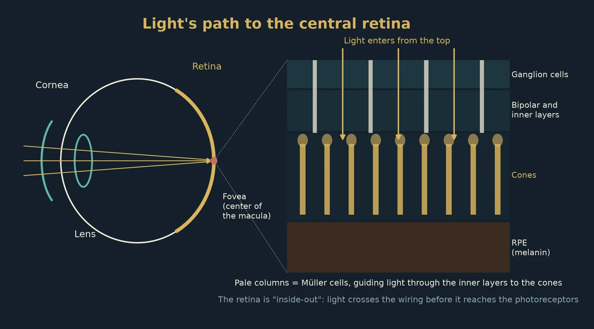
The retina is a thin sheet of neural tissue lining the back of the eye, about a quarter of a millimeter thick in most places. It is wired in a way that surprises most people. The light-detecting cells, the rods and cones, sit at the very back, facing away from the incoming light. Light must first pass through the nerve fiber layer, the ganglion cells, and the layers of bipolar, horizontal and amacrine cells before reaching the photoreceptors. Behind the photoreceptors is the retinal pigment epithelium (RPE), a single layer of dark, melanin-filled cells that absorbs stray light and keeps the photoreceptors alive.
For more than a century this "inverted" design was seen as an optical handicap. Part of the answer to why it works so well came from the glial cells, which is covered below.
The central retina: macula, fovea and foveola
Near the center of the retina is the macula, an oval region about 5 to 6 millimeters across that handles detailed central vision. In its middle is the fovea, a small pit about 1.5 millimeters wide. At the very bottom of the pit is the foveola, roughly 0.35 millimeters wide, which holds only cones, and no rods at all.
At the fovea the inner retinal layers are pushed aside, so light reaches the cones with almost nothing in its way. Christine Curcio and her colleagues counted photoreceptors across whole human retinas in 1990, in the Journal of Comparative Neurology. They found an average of about 4.6 million cones and 92 million rods per eye. Peak foveal cone density averaged about 199,000 cones per square millimeter, and it varied widely from person to person. That packing is what lets you read small print.
Because the cone cells at the center are pushed so close together, their connections can't sit directly beneath them. The cone fibers run sideways, out of the pit, before they reach their partner cells. These sideways fibers are called Henle fibers. They make a ring-shaped layer around the fovea, and they contain much of the macula's yellow pigment.
Macular pigment: lutein, zeaxanthin and meso-zeaxanthin
The macula is yellow. The color comes from three carotenoids that the body gets from food: lutein, zeaxanthin and meso-zeaxanthin, the last of which is formed mostly in the retina from lutein. Richard Bone, John Landrum and their colleagues spent decades mapping these pigments. They found zeaxanthin and meso-zeaxanthin concentrated at the center of the fovea, and lutein spread more widely.
Macular pigment absorbs blue light most strongly, with a peak near 460 nanometers. Researchers describe two main jobs for it. First, it filters short-wavelength light before it reaches the cones, which cuts down blur from chromatic aberration and glare. Second, these carotenoids are antioxidants that quench excited oxygen states in a tissue that is bathed in light and oxygen all day. Large trials such as the AREDS2 study (2013) tested lutein and zeaxanthin in place of beta-carotene in supplements for people with intermediate age-related macular degeneration.
Müller cells: living optical fibers
Müller cells are glial cells that stretch through the full thickness of the retina, from the inner surface to the photoreceptors. In 2007, Kristian Franze, Jochen Guck, Andreas Reichenbach and their colleagues published a study in PNAS titled "Müller cells are living optical fibers in the vertebrate retina."
They showed that Müller cells are funnel-shaped, have a higher refractive index than the tissue around them, and line up along the path of incoming light. Using a dual-beam laser trap, they showed that single Müller cells capture light and guide it end to end, the way a fiber-optic cable does. In their words, "Müller cells seem to mediate the image transfer through the vertebrate retina with minimal distortion and low loss."
Later work by Amichai Labin and Erez Ribak (2010, Physical Review Letters) modeled this light guiding and argued that it helps explain why the inverted retina works so well. In the fovea itself, the inner layers are already pushed aside, so the light path to the central cones is direct.
Photoreceptors: built for catching light
Each rod and cone has two main parts.
- The outer segment is a stack of hundreds of membrane discs (in rods) or folds (in cones) packed with light-sensitive pigment.
- The inner segment holds the cell's machinery. Its outer part, called the ellipsoid, is packed tight with mitochondria.
Photoreceptors are among the most energy-hungry cells in the body. In the dark they keep ion channels open and constantly pump ions back out, a "dark current" that uses a great deal of ATP. That is why the ellipsoid is packed with mitochondria, and it is why the retina is one of the most oxygen-hungry tissues there is.
Cones are also wave guides. Each cone is narrow, has a higher refractive index than its surroundings, and points toward the pupil. In 1933, Walter Stiles and Brian Crawford found that light entering through the center of the pupil looks brighter than the same light entering near the edge. This is now called the Stiles-Crawford effect, and for decades it was explained by the light-guiding shape of the cones. A newer study, described in Pillar 2, links it directly to the mitochondria.
The pigments: rhodopsin and the cone opsins
Rods contain rhodopsin, which absorbs best near 498 nanometers. Cones come in three types, with pigments that peak in the violet-blue (near 420 nm), green (near 530 nm) and yellow-green (near 560 nm). Each pigment is an opsin protein wrapped around the same small molecule, 11-cis retinal, which comes from vitamin A. The protein around it tunes which colors it absorbs.
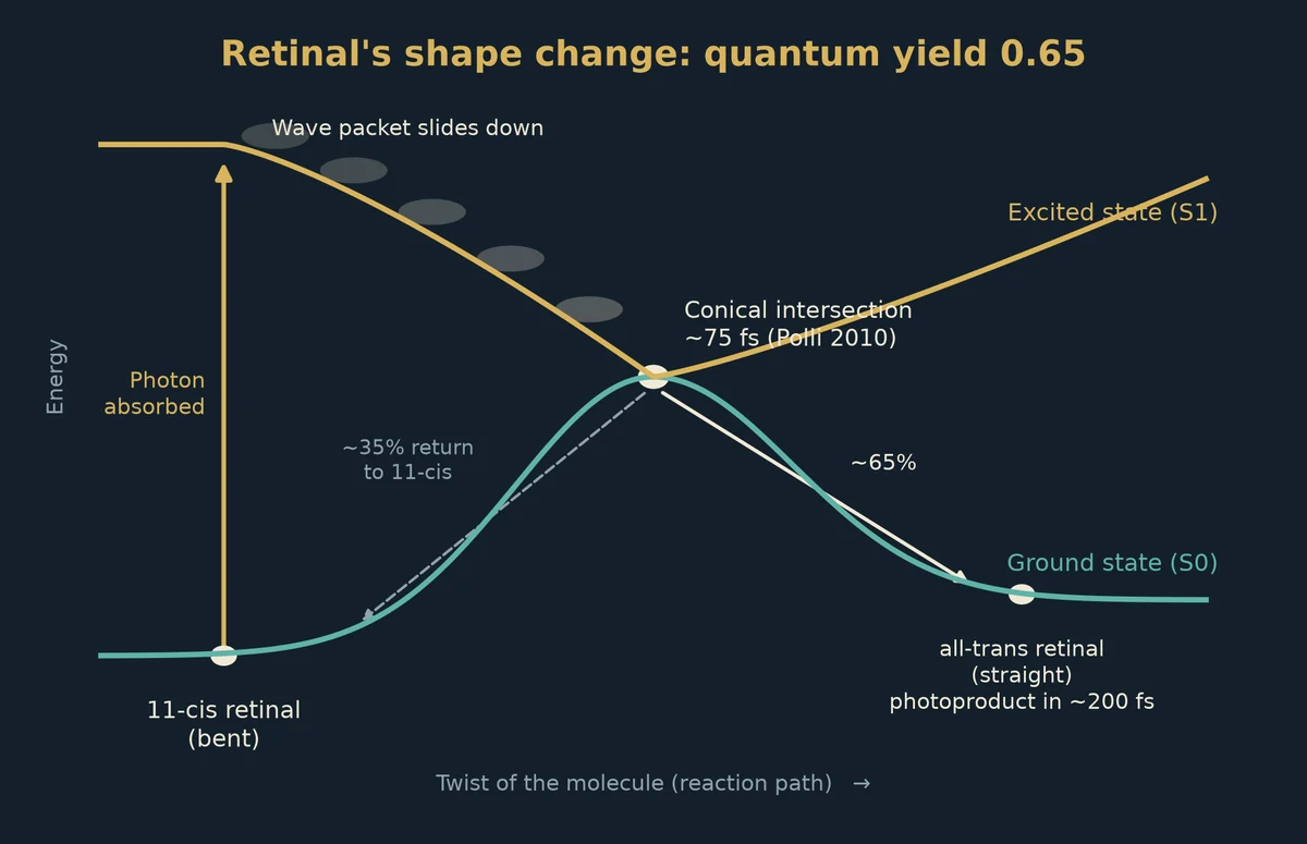
When the retinal absorbs a photon, it twists from its bent 11-cis form into the straight all-trans form. That one change in shape pushes the protein into its active state. Everything else in seeing follows from it.
The first step in vision: a femtosecond quantum event
How fast does that twist happen? In 1991, Robert Schoenlein, Richard Mathies, Charles Shank and their colleagues at Berkeley used femtosecond laser pulses to watch it. In Science, they reported that rhodopsin's first photoproduct appears within about 200 femtoseconds. A femtosecond is a millionth of a billionth of a second. This is one of the fastest chemical reactions known in nature.
The speed is a clue to how it happens. In ordinary chemistry, a molecule gets excited, jiggles around, and eventually finds a way to drop back down. Rhodopsin does something different. Physicists describe the excited molecule as a wave packet, a quantum object whose shape and energy are spread out over many possible arrangements at once. That wave packet slides down the excited-state energy surface and passes through a conical intersection. This is a point where the excited and ground-state energy surfaces touch, forming a funnel that drops the molecule almost directly into its new shape.
In 2010, Dario Polli, Giulio Cerullo, Marco Garavelli, Richard Mathies and their colleagues published "Conical intersection dynamics of the primary photoisomerization event in vision" in Nature. Using laser pulses shorter than 20 femtoseconds, they followed the wave packet moving coherently from the moment of excitation to the photoproduct. Their team's follow-up report puts the arrival at the conical intersection at roughly 75 femtoseconds. Their measurements matched computer simulations that include a true crossing between electronic states. The authors called their findings the most compelling evidence to date that conical intersections matter in visual photochemistry.
The reaction is also efficient. Polli and colleagues cite a quantum yield of 0.65. That means about two out of every three photons that rhodopsin absorbs lead to the switch. This makes vision one of the clearest cases in biology where quantum mechanics is not just a background law but the actual mechanism: the wave-like behavior of the molecule shapes how the reaction turns out.
Counting single photons
Rods are sensitive enough to respond to a single photon. Denis Baylor, Trevor Lamb and King-Wai Yau showed this in toad rods in 1979, recording a small, clear electrical response to single photons. In 1942, Selig Hecht, Simon Shlaer and Maurice Pirenne estimated that a human in the dark could see a flash when only a handful of photons were absorbed. In 2016, Jonathan Tinsley, Alipasha Vaziri and colleagues reported in Nature Communications that people could detect single-photon flashes slightly better than chance, using a light source that emits photons one at a time.
Phototransduction: from one molecule to an electrical signal
Once rhodopsin switches on, a chain reaction amplifies the signal.
1. The active rhodopsin activates many copies of a G-protein called transducin.
2. Transducin switches on an enzyme, phosphodiesterase, that breaks down the messenger molecule cyclic GMP.
3. As cyclic GMP falls, ion channels in the outer segment close.
4. The cell's voltage shifts to more negative values, and it releases less of its chemical messenger, glutamate.
In other words, photoreceptors signal light by going quieter. The used all-trans retinal is passed to the RPE, which turns it back into 11-cis retinal in a cycle called the visual cycle. Cones also have a second recycling route through Müller cells, which helps them recover quickly in bright daylight.
The retinal pigment epithelium and its melanin
Behind the photoreceptors, the RPE does several jobs at once. It recycles retinal, it eats the worn-out tips of the outer segments every day (about a tenth of each outer segment is renewed daily in rods), it supplies nutrients from the blood vessels behind it, and its melanin absorbs light that gets past the photoreceptors. Without that dark layer, light would bounce around inside the eye and blur the image. RPE melanin also binds metals and acts as an antioxidant. Researchers including Tadeusz Sarna have studied how RPE melanin changes with age and how its protective role weakens over time.
The output: bipolar cells and retinal ganglion cells
Photoreceptors pass their signal to bipolar cells, which pass it to retinal ganglion cells (RGCs). Horizontal and amacrine cells shape the signal along the way, sharpening edges and contrast. In the fovea, many cones have a near "private line": one cone connects to one bipolar cell, which connects to one ganglion cell. That one-to-one wiring is why central vision is so sharp. In the outer retina, many rods share a single ganglion cell, which trades detail for sensitivity in dim light.
The ganglion cells are the only cells that send signals out of the eye. Their axons gather into the optic nerve, about a million fibers per eye.
The optic nerve, chiasm and lateral geniculate nucleus
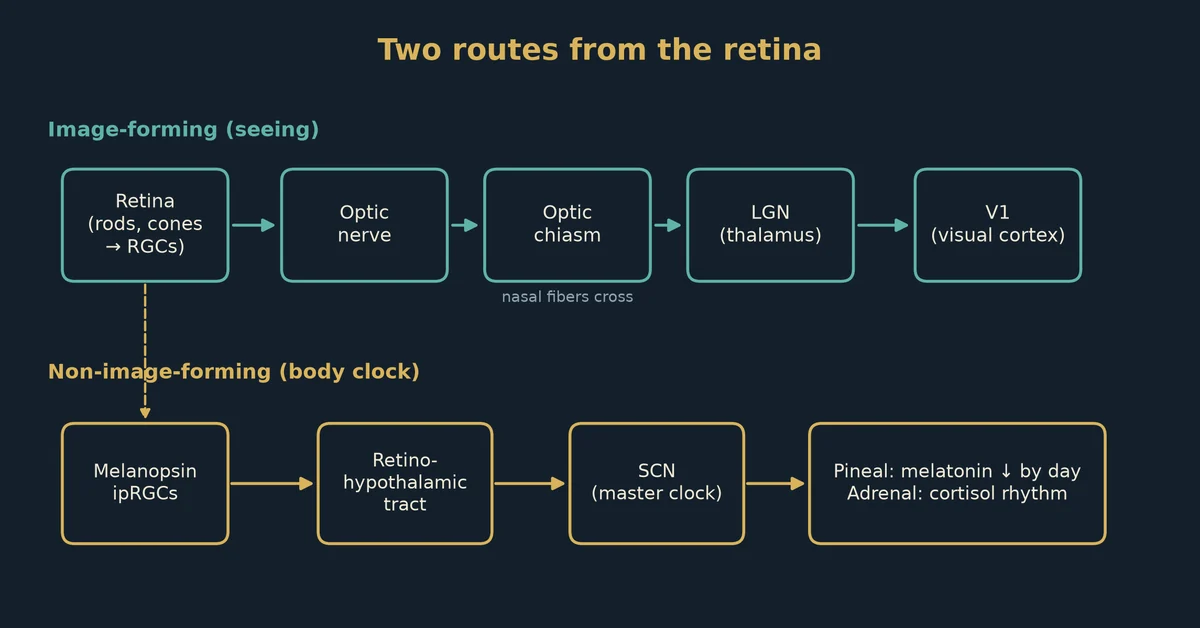
The two optic nerves meet at the optic chiasm, just under the front of the brain. There, fibers from the inner (nasal) half of each retina cross to the other side. As a result, the left half of the brain gets information about the right half of the visual world from both eyes, and vice versa.
Most fibers then travel to the lateral geniculate nucleus (LGN) of the thalamus. The LGN is a relay with six main layers, sorted by eye and cell type: large-cell (magnocellular) layers that carry motion and contrast, small-cell (parvocellular) layers that carry fine detail and red-green color, and thin koniocellular layers in between that carry some blue-yellow signals.
Primary visual cortex (V1)
From the LGN, a fan of fibers called the optic radiation carries the signal to the primary visual cortex, V1, at the back of the brain. David Hubel and Torsten Wiesel won the 1981 Nobel Prize for showing how V1 neurons respond to lines and edges at particular angles. The fovea takes up only a tiny part of the retina, but it claims a large share of V1. This is called cortical magnification, and it is why the center of your gaze feels so rich in detail.
The second pathway: melanopsin, ipRGCs and the body clock
For most of the twentieth century, scientists assumed rods and cones were the only light sensors in the eye. That changed around 2000.
- In 1998, Ignacio Provencio and colleagues found a new light-sensitive pigment, melanopsin, first in frog skin and then in the mammalian retina.
- In 2002, David Berson, Felice Dunn and Motoharu Takao showed in Science that a small group of ganglion cells in the rat retina respond to light on their own, even when cut off from rods and cones. In the same issue, Samer Hattar, King-Wai Yau and colleagues showed that these cells contain melanopsin and send their axons to the brain's master clock.
These cells are now called intrinsically photosensitive retinal ganglion cells (ipRGCs). They make up only a few percent of ganglion cells. Melanopsin responds best to blue light near 480 nanometers. The cells respond slowly and keep firing as long as the light lasts, which makes them good at measuring overall brightness rather than detail.
The ipRGCs send their axons along the retinohypothalamic tract to the suprachiasmatic nucleus (SCN), a pair of tiny nuclei just above the optic chiasm. The SCN is the body's master clock. Light in the morning moves the clock earlier, and light late in the evening moves it later. ipRGCs also drive the pupil's response to light and connect to brain areas involved in sleep, alertness and mood.
Downstream: melatonin, cortisol and the rest of the body
From the SCN, signals pass through a chain of nerves to the pineal gland, which makes melatonin at night. In 1980, Alfred Lewy and colleagues showed in Science that bright light suppresses melatonin in humans. Later studies by George Brainard and colleagues (2001) and Kavita Thapan and colleagues (2001) found that the strongest melatonin suppression comes from blue light, which pointed to a non-rod, non-cone sensor before melanopsin was confirmed in humans.
The SCN also sets the timing of the daily cortisol rise in the morning, body temperature, and the clocks found in nearly every organ. Joseph Takahashi, Ueli Schibler and others showed that cells in the liver, muscle and fat have their own clock genes, and that the SCN keeps them in step. These peripheral clocks control the timing of metabolism, including when mitochondria are most active. In this way, light falling on a few thousand cells in the eye reaches the whole body.
Pillar 2: Middle Ground: active and contested research
Middle Ground
Mitochondria as microlenses
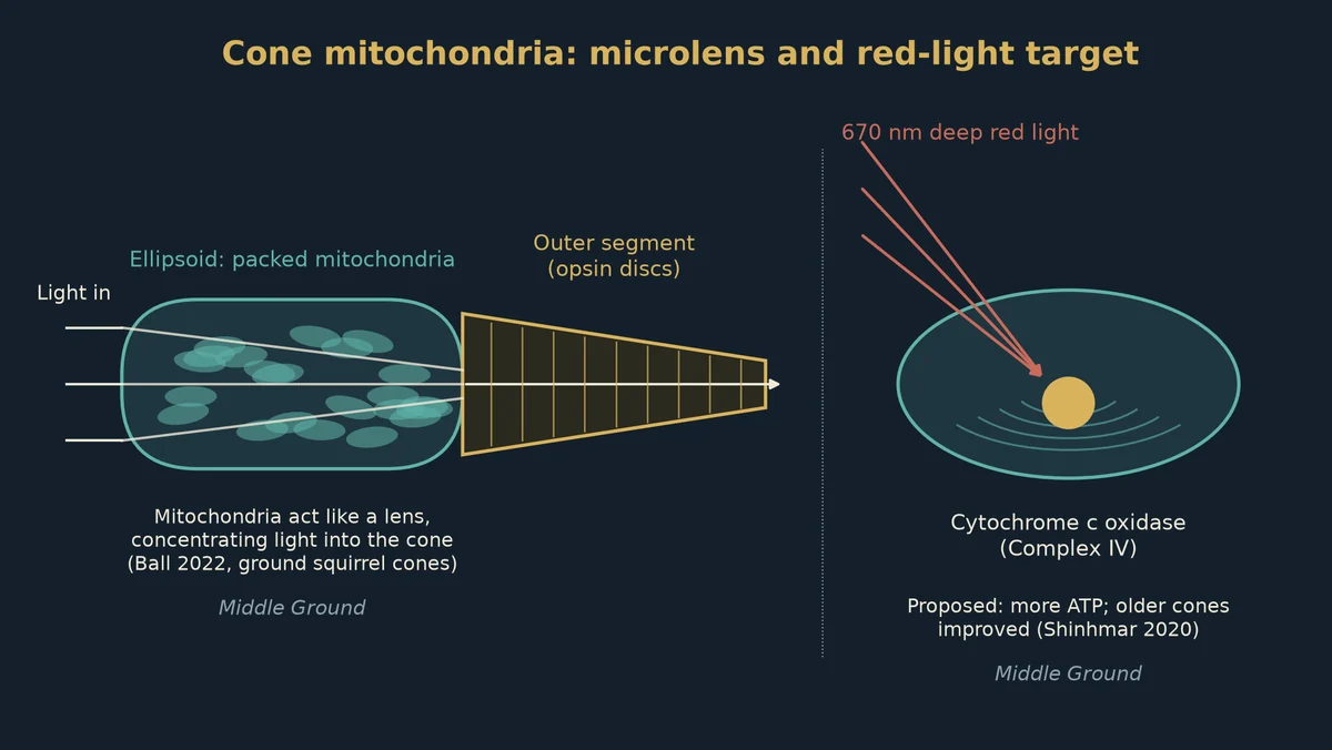
In 2022, John Ball, Wei Li and their colleagues at the U.S. National Eye Institute published "Mitochondria in cone photoreceptors act as microlenses to enhance photon delivery and confer directional sensitivity to light" in Science Advances.
They studied the thirteen-lined ground squirrel, which has a retina dominated by cones and which hibernates. Using live imaging and computer modeling of real mitochondrial shapes, they showed that the tightly packed mitochondria in the cone ellipsoid focus light into a thin beam aimed at the outer segment. They wrote that "far from being detrimental to light detection, cone mitochondria alone are sufficient to focus incident light" to a thin pencil of light. The light delivered this way had the same direction dependence as the Stiles-Crawford effect, which offers a new explanation for that old finding.
They also compared active and hibernating squirrels. In hibernating animals, whose mitochondria are less orderly, focusing was weaker. Light concentrated in the outer-segment region was about 22 percent higher in active animals. The team noted that how well mitochondria focus light depends on their refractive index, which can change with their respiration state.
Why is this Middle Ground and not Centralized? The work is strong and peer-reviewed, but it is one research group's study in one animal. Whether the same thing happens in human cones, and how much it matters for everyday vision, is still being tested.
Red light, aging cones and cytochrome c oxidase
Glen Jeffery at University College London has spent years studying how long-wavelength light affects aging retinas. His lab and others report that deep red and near-infrared light (roughly 650 to 1,000 nanometers) is absorbed by mitochondria, especially by cytochrome c oxidase, the last enzyme in the chain that makes ATP. This field is called photobiomodulation. Tiina Karu's work in Russia in the 1980s and 1990s first proposed cytochrome c oxidase as the main red-light absorber.
Jeffery's group first tested deep red light in mice, bumblebees and fruit flies, and reported better mitochondrial function and retinal performance in older animals. In 2020, Harpreet Shinhmar, Glen Jeffery and colleagues published "Optically Improved Mitochondrial Function Redeems Aged Human Visual Decline" in the Journals of Gerontology.
- Who: 24 healthy adults aged 28 to 72 with no eye disease.
- What: each looked into a small 670-nanometer deep red light for 3 minutes a day for two weeks.
- Results: in people around 40 and older, color contrast sensitivity on the blue (tritan) axis improved significantly, by about 22 percent on average in those over 38. The red (protan) axis improved by about 10 percent, but not significantly. Rod sensitivity in the dark also improved significantly in those over 40. Younger people showed no change.
In the study's abstract, the authors note that photoreceptors have the highest mitochondrial density in the body and that the retina loses about 70 percent of its ATP over a lifetime. In UCL's press release, Jeffery described the effect as using light wavelengths that recharge the retina's declining energy system, "rather like re-charging a battery."
The group later reported that timing matters. In a 2021 study in Scientific Reports, a single 3-minute morning exposure to 670-nanometer light improved color contrast for about a week in older people, and the same exposure in the afternoon had no effect. They linked this to daily rhythms in mitochondrial activity.
Why Middle Ground? The studies are small, and the exact absorbing molecule and mechanism are still debated. Some scientists question whether cytochrome c oxidase is the main target, and others point to water or other molecules near mitochondria. Red-light devices for dry macular degeneration have been tested in clinical trials (such as the LIGHTSITE trials), with mixed reactions from eye specialists. People should not stare into lasers or unregulated devices; the studies used specific, low-power lights.
Macular pigment beyond filtering
Some research groups, including John Nolan and Stephen Beatty in Ireland and Billy Hammond in the United States, have studied whether higher macular pigment levels improve contrast, glare recovery and even processing speed. Their trials report modest benefits from lutein and zeaxanthin supplements in some measures. Other researchers find the effects small or inconsistent. The protective filtering role is Centralized; the performance benefits sit in the Middle Ground.
The radical-pair compass: cryptochromes in the retina
Many migratory birds can sense the direction of the Earth's magnetic field, and for some species this sense depends on light. Wolfgang and Roswitha Wiltschko showed in the 1970s and later that robins orient by the field's inclination and need short-wavelength light to do it.
In 2000, Thorsten Ritz, Salih Adem and Klaus Schulten proposed in the Biophysical Journal that birds sense the field through a radical pair. In this model, light makes a protein in the retina transfer an electron, which creates two molecules with unpaired electrons whose spins are linked. The Earth's weak magnetic field nudges how those spins evolve, which changes how much of a chemical product forms. This is a quantum effect involving electron spin. The leading candidate protein is cryptochrome, found in the retina. In 2021, Jingjing Xu, Henrik Mouritsen, Peter Hore and colleagues reported in Nature that robin cryptochrome 4 is magnetically sensitive in the lab. For birds, the radical-pair idea is now strongly supported, though not every detail is settled.
Humans also have cryptochromes. In 2011, Lauren Foley, Robert Gegear and Steven Reppert at the University of Massachusetts put the human CRY2 gene into fruit flies that lacked their own cryptochrome. The flies regained a light-dependent response to magnetic fields, and it needed blue light. The authors wrote that "human CRY2 has the molecular capability to function as a light-sensitive magnetosensor." They were careful to say this shows the protein's capability, not that humans actually sense magnetic fields.
In 2019, Connie Wang, Joseph Kirschvink, Shinsuke Shimojo and colleagues published an EEG study in eNeuro. They reported that some people's brain alpha waves dropped in a repeatable way when an Earth-strength magnetic field was rotated around them, but only when the field pointed downward, as it does in the Northern Hemisphere. Because the response depended on the field's polarity, the authors argued against the cryptochrome model: "This rules out free-radical 'quantum compass' mechanisms like the cryptochrome hypothesis, which can detect only axial alignment." They favored a magnetite-based mechanism instead. Their findings have not yet been widely replicated.
So, for humans, the picture is split. Cryptochrome sits in the retina and can work as a magnetic sensor in flies. One EEG study found a brain response but pointed away from cryptochrome. Whether people have any working magnetic sense, and whether it runs through the retina, is still an open question.
Light made inside the eye: ultraweak photon emission
All living tissue gives off a very faint glow of light, called ultraweak photon emission (UPE) or biophotons, mostly from oxidation reactions. In 2011, Zhuo Wang, István Bókkon, Jiapei Dai and Ildikó Antal reported in Brain Research that freshly isolated rat eyes, lenses, vitreous and retinas give off ultraweak photons on their own, and give off more after being exposed to visible light.
Bókkon and his colleagues proposed that this internal light could explain some phosphenes, the flashes and specks people see with their eyes closed or in total darkness. In their view, light made by oxidation inside the retina could be absorbed by the photoreceptors and read as light. This proposal is plausible on physics grounds, because rods can respond to single photons, but direct proof in living human eyes is still missing.
Pillar 3: Decentralized View: ideas not yet established
Decentralized View
Light traveling inside nerves
In 2010, Yan Sun, Chao Wang and Jiapei Dai published "Biophotons as neural communication signals demonstrated by in situ biophoton autography" in Photochemical & Photobiological Sciences. They shone light on one end of rat spinal nerve roots and reported more biophoton activity at the other end, which they read as light being carried along the nerve. In 2016, Sourabh Kumar, Kristine Boone, Jack Tuszyński, Paul Barclay and Christoph Simon published a modeling study in Scientific Reports titled "Possible existence of optical communication channels in the brain." They calculated that the myelin wrapped around axons could, in principle, guide light like a fiber, and they proposed experiments to test it.
The researchers in this field describe a possible second, optical channel running alongside the electrical signals in nerves. The optic nerve, made of about a million heavily myelinated fibers, is one of the places they point to. Supporters say the eye, which already uses light and carries Müller-cell and mitochondrial light guides, would be a natural place for such a system. The researchers themselves stress that the modeling shows possibility, not proof, and no one has shown that the brain uses such light as information in a living animal.
Persinger and light in the brain
The late Michael Persinger at Laurentian University in Canada and his colleagues reported in the 2010s that photon emission from people's heads increased when they imagined light, and they linked these changes to magnetic fields and to brain activity. Persinger argued that biophotons and weak magnetic fields could be part of how the brain stores and shares information. His work drew interest and strong criticism, because the results were small, hard to repeat and measured with methods other labs questioned.
Bókkon's "visual imagery as biophysical pictures"
István Bókkon has proposed that biophotons made inside the visual cortex may help create mental images, so that imagining something involves real, faint light in the brain's visual areas. He describes the retina-to-V1 pathway as possibly carrying both electrical and optical signals. This idea has not been tested directly.
Mitochondria, light and the "inner sun"
Some writers and clinicians, including the neurosurgeon Dr. Jack Kruse, describe the retina as the body's main light sensor for health, not just for sight. In Kruse's framing, natural sunlight through the eyes, especially at sunrise, sets the circadian clock, tunes mitochondria throughout the body, and supports hormones such as melatonin and dopamine. He also argues that blue-heavy artificial light at night disrupts this system, and that melanin and DHA in the retina play special roles in capturing and using light energy. His views draw from real findings (the ipRGC-SCN pathway, red light and mitochondria, the high mitochondrial density of photoreceptors), but many of his larger claims, such as specific quantum effects of DHA or water in the retina, have not been tested in published studies.
DHA and the photoreceptor membrane
Photoreceptor outer segments contain some of the highest levels of the omega-3 fat DHA in the body. The Centralized View holds that DHA keeps disc membranes flexible, which helps rhodopsin change shape quickly. Michael Crawford and colleagues have argued that DHA was essential for the evolution of the vertebrate eye and brain, and that its many double bonds may help with fast electron movement and signaling. The structural role is established. The proposed quantum or electron-transfer roles of DHA in vision are Decentralized View.
The retina as a magnetic sensor in humans
Some writers go further than the published evidence and describe human eyes as active magnetic sensors that tie people to the Earth's field and to the Sun's activity. As covered in Pillar 2, the published human evidence is limited to the fruit-fly CRY2 study and one EEG study, and that EEG study pointed away from a retina-based quantum compass. Claims of a working human retinal magnetic sense belong here.
The Retina and the Leptin-Melanocortin Pathway
This section looks at a second body system that light from the eye may reach: the brain's appetite and energy-balance circuit. Each claim is labeled with the view it belongs to.
The appetite circuit itself Centralized View
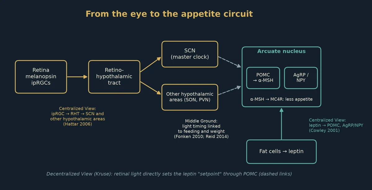
In 1994, Yiying Zhang, Jeffrey Friedman and colleagues at Rockefeller University reported in Nature the cloning of the ob gene, which codes for leptin, a hormone made mainly by fat cells. The more fat the body stores, the more leptin it releases. Leptin travels to the arcuate nucleus at the base of the hypothalamus, where it acts on two groups of neurons with opposite jobs:
- POMC neurons, which make proopiomelanocortin. POMC is cut into several smaller peptides, including α-MSH, ACTH and β-endorphin. α-MSH lowers appetite and raises energy use.
- AgRP/NPY neurons, which make agouti-related peptide and neuropeptide Y. These raise appetite and lower energy use.
In 2001, Michael Cowley, Roger Cone, Malcolm Low and colleagues showed in Nature that leptin increases the firing of POMC neurons in mice in two ways: it excites them directly, and it reduces the inhibitory input they receive from nearby NPY/GABA neurons. α-MSH acts on the melanocortin-4 receptor (MC4R) and the related MC3R. In 1997, Dennis Huszar, Roger Cone and colleagues reported in Cell that mice lacking MC4R overeat and become obese; by 15 weeks, females were about twice as heavy as normal littermates and males about 50% heavier. The same POMC-derived α-MSH, acting on a different receptor (MC1R) in pigment cells, drives the production of dark melanin in skin and hair.
How light from the eye reaches the hypothalamus Centralized View
The ipRGC pathway described in Pillar 1 does not stop at the SCN. In 2006, Samer Hattar, King-Wai Yau and colleagues mapped where melanopsin cells send their fibers in mice (Journal of Comparative Neurology). Besides the SCN, they found projections to several other hypothalamic areas, the preoptic area, the medial amygdala, the lateral habenula and other brain regions. Through the SCN, light also sets the timing of the clocks in fat, liver and muscle, and of feeding and leptin release.
In 2023, Jian Jun Meng and colleagues in Tian Xue's lab at the University of Science and Technology of China reported in Cell that light worsens glucose tolerance in mice through a route that starts with melanopsin ipRGCs. The signal runs to the supraoptic nucleus of the hypothalamus (not the SCN), then to the paraventricular nucleus and on to the brainstem, and it lowers heat production by brown fat. The effect disappeared in mice without melanopsin. The team also found that in people, light reduced glucose tolerance in conditions where brown fat was active, but not at a warm, neutral temperature. This is a published retina-to-hypothalamus-to-metabolism route, though it runs through the supraoptic and paraventricular nuclei rather than directly through the arcuate POMC neurons.
Leptin and melanocortins inside the eye Centralized View
The eye has its own pieces of this system:
- α-MSH in the eye. In 1992, Andrew Taylor, J. Wayne Streilein and Scott Cousins reported in Current Eye Research that α-MSH is present in normal aqueous humor of humans (about 20 picomolar), rabbits and mice, at levels that suppress the inflammatory signal interferon-gamma from activated immune cells. Taylor's group has since described α-MSH as one of the factors behind the eye's immune privilege, its ability to limit inflammation.
- Melanocortins and retinal survival. In 2003, Nava Naveh reported in Graefe's Archive for Clinical and Experimental Ophthalmology that melanocortins injected into the eye delayed retinal degeneration in Royal College of Surgeons rats, a strain with inherited retinal dystrophy.
- Leptin in the retina. In 2004, Eri Suganami, Hitoshi Takagi, Nagahisa Yoshimura and colleagues reported in Diabetes that leptin promotes the growth of new blood vessels in the oxygen-starved mouse retina, partly through VEGF, and that the leptin receptor is found on retinal blood-vessel cells. Other teams have reported leptin receptors in retinal astrocytes and Müller glia. Broad leptin-receptor signaling in the retina's light-sensing neurons has not been shown.
Light timing, weight and leptin Middle Ground
- Light at night in mice. In 2010, Laura Fonken, Randy Nelson and colleagues at Ohio State University reported in PNAS that mice kept in dim light at night (about 5 lux) gained more weight and fat and handled glucose worse than mice in normal light and dark, even though they ate the same total amount of food. The dim-light mice ate 55.5% of their food during the light phase, versus 36.5% in normal-cycle mice. When their food was limited to the active dark phase, the extra weight gain did not occur. The authors concluded that light at night shifted the timing of eating.
- Morning light in people. In 2014, Kathryn Reid, Phyllis Zee and colleagues at Northwestern University reported in PLOS ONE that among 54 adults who wore light sensors for a week, those whose bright light (above 500 lux) came earlier in the day had a lower body mass index. Each hour later was linked to about 1.28 more BMI units, independent of sleep timing, activity, age and season. The study shows a link, not cause and effect. Reid said: "The earlier this light exposure occurred during the day, the lower the individuals' body mass index."
- Mice without melanopsin. A 2015 PLOS ONE study reported that melanopsin-knockout mice lost much more weight than normal mice on a ketogenic diet, while food intake and activity looked similar early on. How melanopsin affects metabolism in these mice is still unclear.
These findings tie the eye's light signals to eating patterns and weight. They do not yet show that the retina's signals change leptin action or POMC firing in the arcuate nucleus directly.
One light-to-metabolism axis Decentralized View
Some writers join these pieces into a single system. The most prominent is the neurosurgeon Dr. Jack Kruse, whose 2018 blog post "QT #22: Leptin Resistance is Melanopsin Dysfunction" sets out the idea in its title. In his framing:
- Leptin resistance is, in his words, "a photonic process in human biology." He describes leptin as the hormone that controls energy metabolism, and ties it to light signals from the eye and skin through the body's circadian clocks.
- Because one gene, POMC, makes both α-MSH (appetite control and melanin) and ACTH and β-endorphin (stress and mood), writers in this camp, Kruse among them, treat light, melanin, hormones and metabolism as one connected system.
- He argues that screens, blue light and non-native electromagnetic fields disrupt the bond between melanopsin and its vitamin A partner, which he says leads to leptin resistance and hunger. He links this to DHA in cell membranes and to mitochondrial function.
Writers in the quantum-biology community extend this further, suggesting that ultraweak photons, melanin and mitochondria in the retina form part of a light-to-fat signaling chain. None of these links has been tested as a single pathway in published studies. The parts that are established are the separate pieces: the ipRGC-to-hypothalamus routes, the leptin-POMC-MC4R circuit, POMC's role in melanin, and the eye's own α-MSH. The claim that retinal light sets leptin sensitivity through POMC is Decentralized View.
The whole route at a glance
| Stage | What happens | Level |
|---|---|---|
| Cornea and lens | Focus light, filter UV | Centralized View |
| Macular pigment | Filters blue light, antioxidant | Centralized View |
| Müller cells | Guide light to the photoreceptors | Centralized View (Franze 2007) |
| Cone mitochondria | Focus light into the outer segment | Middle Ground (Ball 2022) |
| Retinal isomerization | Reaches the conical intersection in about 75 fs; all-trans retinal appears within about 200 fs | Centralized View |
| Phototransduction | Amplifies one photon into an electrical signal | Centralized View |
| RGCs, chiasm, LGN, V1 | Carry and build the image | Centralized View |
| ipRGCs, RHT, SCN | Set the body clock, melatonin, cortisol | Centralized View |
| Red light and mitochondria | Improves aging cone function | Middle Ground (Shinhmar 2020) |
| Cryptochrome compass | Strong in birds, unclear in humans | Middle Ground |
| UPE in the eye | Measured in rat eyes | Middle Ground (Wang 2011) |
| Light signals in nerves | Proposed optical channel | Decentralized View |
| ipRGCs to hypothalamus | Light reaches areas beyond the SCN; glucose route via the supraoptic nucleus | Centralized View (Hattar 2006; Xue 2023) |
| Leptin, POMC, AgRP, MC4R | Arcuate appetite and energy-balance circuit | Centralized View (Cowley 2001; Huszar 1997) |
| α-MSH in the eye | Immune privilege, retinal protection | Centralized View (Taylor 1992) |
| Light timing and weight | Night light and late light linked to weight gain | Middle Ground (Fonken 2010; Reid 2014) |
| Light-set leptin "setpoint" | One retina-POMC-metabolism axis | Decentralized View (Kruse) |
Not yet verified
These points have not been established and should be read as open questions:
- That mitochondria focus light into human cones as they do in ground squirrels.
- The exact molecule that absorbs 670-nanometer light in the retina, and how long and how widely the red-light benefits last.
- That red-light devices are a proven treatment for macular degeneration.
- That humans have a working magnetic sense, or that one runs through retinal cryptochrome.
- That ultraweak photons made inside the eye cause phosphenes in living people.
- That myelinated axons in the optic nerve or brain carry light as a signal.
- That the visual cortex uses biophotons to make mental images.
- That DHA in the retina has a special quantum or electron-transfer role in vision.
- Persinger's reports linking imagined light, photon emission and magnetic fields.
- That retinal light signals act directly on arcuate POMC or AgRP/NPY neurons, or set leptin sensitivity.
- That leptin resistance is caused by melanopsin dysfunction or artificial light at night.
- That the light-timing and weight links in mice and people are cause and effect through the leptin-melanocortin pathway.
- That retinal melanin, ultraweak photons or mitochondria feed a single light-to-fat chain through POMC.
- How melanopsin loss changes weight in mice on a ketogenic diet.
- Broad claims that sunlight through the eyes "tunes" every mitochondrion in the body, beyond the established clock pathway.
This article presents viewpoints, attributed to their sources. It is not medical advice.
Key sources
- Curcio CA et al. (1990). Human photoreceptor topography. Journal of Comparative Neurology 292:497–523.
- Franze K et al. (2007). Müller cells are living optical fibers in the vertebrate retina. PNAS 104:8287–8292. https://doi.org/10.1073/pnas.0611180104
- Labin AM, Ribak EN (2010). Retinal glial cells enhance human vision acuity. Physical Review Letters 104:158102.
- Schoenlein RW et al. (1991). The first step in vision: femtosecond isomerization of rhodopsin. Science 254:412–415. https://doi.org/10.1126/science.1925597
- Polli D et al. (2010). Conical intersection dynamics of the primary photoisomerization event in vision. Nature 467:440–443. https://doi.org/10.1038/nature09346
- Baylor DA, Lamb TD, Yau KW (1979). Responses of retinal rods to single photons. Journal of Physiology 288:613–634.
- Tinsley JN et al. (2016). Direct detection of a single photon by humans. Nature Communications 7:12172.
- Berson DM, Dunn FA, Takao M (2002). Phototransduction by retinal ganglion cells that set the circadian clock. Science 295:1070–1073.
- Hattar S et al. (2002). Melanopsin-containing retinal ganglion cells. Science 295:1065–1070.
- Lewy AJ et al. (1980). Light suppresses melatonin secretion in humans. Science 210:1267–1269.
- Brainard GC et al. (2001). Journal of Neuroscience 21:6405–6412. Thapan K et al. (2001). Journal of Physiology 535:261–267.
- Ball JM et al. (2022). Mitochondria in cone photoreceptors act as microlenses. Science Advances 8:eabn2070. https://doi.org/10.1126/sciadv.abn2070
- Shinhmar H et al. (2020). Optically improved mitochondrial function redeems aged human visual decline. Journals of Gerontology A. https://doi.org/10.1093/gerona/glaa155
- Shinhmar H et al. (2021). Weeklong improved colour contrasts sensitivity after single 670 nm exposures. Scientific Reports.
- Ritz T, Adem S, Schulten K (2000). A model for photoreceptor-based magnetoreception in birds. Biophysical Journal 78:707–718.
- Xu J et al. (2021). Magnetic sensitivity of cryptochrome 4 from a migratory songbird. Nature 594:535–540.
- Foley LE, Gegear RJ, Reppert SM (2011). Human cryptochrome exhibits light-dependent magnetosensitivity. Nature Communications 2:356.
- Wang CX et al. (2019). Transduction of the geomagnetic field as evidenced from alpha-band activity in the human brain. eNeuro 6:ENEURO.0483-18.2019.
- Wang Z, Bókkon I, Dai J, Antal I (2011). Spontaneous and visible light-induced ultraweak photon emission from rat eyes. Brain Research 1369:1–9. https://doi.org/10.1016/j.brainres.2010.10.077
- Sun Y, Wang C, Dai J (2010). Biophotons as neural communication signals demonstrated by in situ biophoton autography. Photochemical & Photobiological Sciences 9:315–322. https://doi.org/10.1039/b9pp00125e
- Kumar S et al. (2016). Possible existence of optical communication channels in the brain. Scientific Reports 6:36508.
- Zhang Y, Proenca R, Maffei M, Barone M, Leopold L, Friedman JM (1994). Positional cloning of the mouse obese gene and its human homologue. Nature 372:425–432.
- Huszar D et al. (1997). Targeted disruption of the melanocortin-4 receptor results in obesity in mice. Cell 88:131–141. https://doi.org/10.1016/S0092-8674(00)81865-6
- Cowley MA et al. (2001). Leptin activates anorexigenic POMC neurons through a neural network in the arcuate nucleus. Nature 411:480–484.
- Hattar S et al. (2006). Central projections of melanopsin-expressing retinal ganglion cells in the mouse. Journal of Comparative Neurology 497:326–349. https://doi.org/10.1002/cne.20970
- Meng JJ, Shen JW, ... Xue T (2023). Light modulates glucose metabolism by a retina-hypothalamus-brown adipose tissue axis. Cell 186:398–412. https://doi.org/10.1016/j.cell.2022.12.024 (PubMed 36669474)
- Taylor AW, Streilein JW, Cousins SW (1992). Identification of alpha-melanocyte stimulating hormone as a potential immunosuppressive factor in aqueous humor. Current Eye Research. https://doi.org/10.3109/02713689208999545
- Naveh N (2003). Melanocortins applied intravitreally delay retinal dystrophy in Royal College of Surgeons rats. Graefe's Archive for Clinical and Experimental Ophthalmology 241:1044–1050.
- Suganami E et al. (2004). Leptin stimulates ischemia-induced retinal neovascularization. Diabetes 53:2443. https://doi.org/10.2337/diabetes.53.9.2443
- Fonken LK et al. (2010). Light at night increases body mass by shifting the time of food intake. PNAS. https://doi.org/10.1073/pnas.1008734107
- Reid KJ et al. (2014). Timing and intensity of light correlate with body weight in adults. PLOS ONE. https://doi.org/10.1371/journal.pone.0092251
- Lack of melanopsin is associated with extreme weight loss in mice upon dietary challenge (2015). PLOS ONE. https://doi.org/10.1371/journal.pone.0127031
- Kruse J (2018). QT #22: Leptin Resistance is Melanopsin Dysfunction. https://jackkruse.com/qt-22-leptin-resistance-is-melanopsin-dysfunction/
- Kruse J. Leptin: Chapter One. https://jackkruse.com/chapter-one-on-leptin/
This article is for education only and is not medical advice. Talk with an eye doctor or other qualified clinician before starting any light therapy, supplement, or change in care, and never look directly at the Sun, lasers, or unregulated light devices.
Better signals, less noise.
One email a week: the best threads on light, Earth cycles and sovereignty, with context, evidence labels and links to the original sources.
Thanks! You're on the list.
Sorry, something went wrong. Please try again, or reach us on X at @primal_buddha.
No spam. Unsubscribe anytime.