 Primal Adapted
The Dark Battery: Melanin's Quantum Biophysics, From Pigment to Radical to Radiation
You've heard of melanin, but why is it so important? It shields against UV, sits in the eye, inner ear and brain, carries stable free radicals and turns nearly all the light it absorbs into heat, which makes it a subject for quantum biophysics.
You've heard of melanin, but why is it so important? Most people know it as the pigment that colors skin, hair and eyes, and as the reason skin tans in summer. That is only the start of what it does.
Melanin is the body's built-in sunscreen. It absorbs ultraviolet and visible light across the whole range, and in laboratory measurements it turns more than 99.9 percent of the light energy it absorbs into heat rather than re-emitting it. It is not only in the skin. It lines the back of the eye, sits in the inner ear and builds up with age in the substantia nigra, the dopamine-making region of the brain that is affected in Parkinson's disease. It carries stable free radicals, unpaired electrons that physicists detect with a magnet and microwaves. It binds metals such as iron and copper. And on the walls of the damaged Chernobyl reactor, scientists found fungi rich in melanin; in later lab studies, melanized fungi grew faster when exposed to ionizing radiation.
Each of those traits comes down to how light, electrons and spin behave inside a disordered pigment, which is why melanin has become a subject for quantum biophysics as well as biology. Researchers agree on some of the story and argue about the rest: whether melanin is a semiconductor, how it carries current, and whether it can capture energy for living things. This article follows that story through the three labels used across the site:
- Centralized View covers what is well established: the chemistry, optics and biology confirmed many times over.
- Middle Ground covers active research that is real and published but still contested.
- Decentralized View covers emerging and unestablished ideas, including melanin as an energy transducer and Dr. Jack Kruse's account of melanin, light and mitochondria.
Each idea sits in the pillar that fits it best. Where a claim has not been shown by research, the text says so.
Pillar 1: Centralized View: the established science
Centralized View
A pigment with a long chemical history
The modern chemistry of melanin begins with an enzyme. In 1927, Henry Stanley Raper at the University of Manchester published "The tyrosinase-tyrosine reaction" in the Biochemical Journal, tracing how the enzyme tyrosinase turns the amino acid tyrosine, step by step, into dark pigment. Through DOPA, dopaquinone and a ring-closing step, the route leads to small building blocks called indoles. Howard Mason later extended the scheme, and the route is often called the Raper–Mason pathway.
What happens after the indoles proved much harder. Melanin does not dissolve, does not crystallize and does not break cleanly into its parts, so the usual tools of chemistry slide off it. In Naples, Rodolfo Nicolaus and his colleagues spent decades degrading natural melanins and reading the fragments, beginning a series of Tetrahedron papers titled "The structure of melanins and melanogenesis" in 1961. Their work, carried on by Giuseppe Prota (who reviewed the field in Medicinal Research Reviews in 1988) and later by Marco d'Ischia and Alessandro Pezzella, established that melanin is not one molecule but a family of irregular polymers and aggregates built from a few kinds of units.
Three melanins
Researchers now group the natural pigments into three main kinds:
- Eumelanin, brown to black, built mainly from 5,6-dihydroxyindole (DHI) and its carboxylic acid cousin DHICA. It dominates dark hair, dark skin and the iris.
- Pheomelanin, red to yellow, built when the amino acid cysteine joins the pathway and sulfur-containing benzothiazine units form. It colors red hair and freckles.
- Neuromelanin, a dark pigment that builds up with age inside certain brain neurons, especially the dopamine neurons of the substantia nigra and the noradrenaline neurons of the locus coeruleus. It is a mixture of melanin, protein, lipid and bound metals.
Shosuke Ito and Kazumasa Wakamatsu of Fujita Health University developed the chemical degradation methods that let labs measure eumelanin and pheomelanin separately in hair, skin and tissue. In their 2003 comparative review in Pigment Cell Research, they showed that most natural pigments are mixtures of the two, in ratios that vary with species, skin type and body site. In 2013, d'Ischia led an international group in Pigment Cell & Melanoma Research that set out shared methods, standards and names for the field, a sign of how hard it had been even to agree on what was being measured.
Broadband absorption: a black body for life
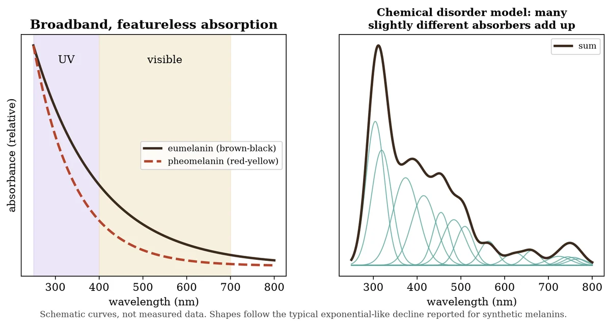
Melanin's best-known job is shielding. Eumelanin absorbs across the whole ultraviolet and visible range, strongest in the UV and fading smoothly toward the red, with none of the sharp peaks that most pigments show. Paul Meredith (then at the University of Queensland) and Tadeusz Sarna (Jagiellonian University, Kraków), in their 2006 review "The physical and chemical properties of eumelanin," describe this smooth, featureless curve as one of the pigment's defining traits, and one that had long resisted explanation.
Light in, heat out
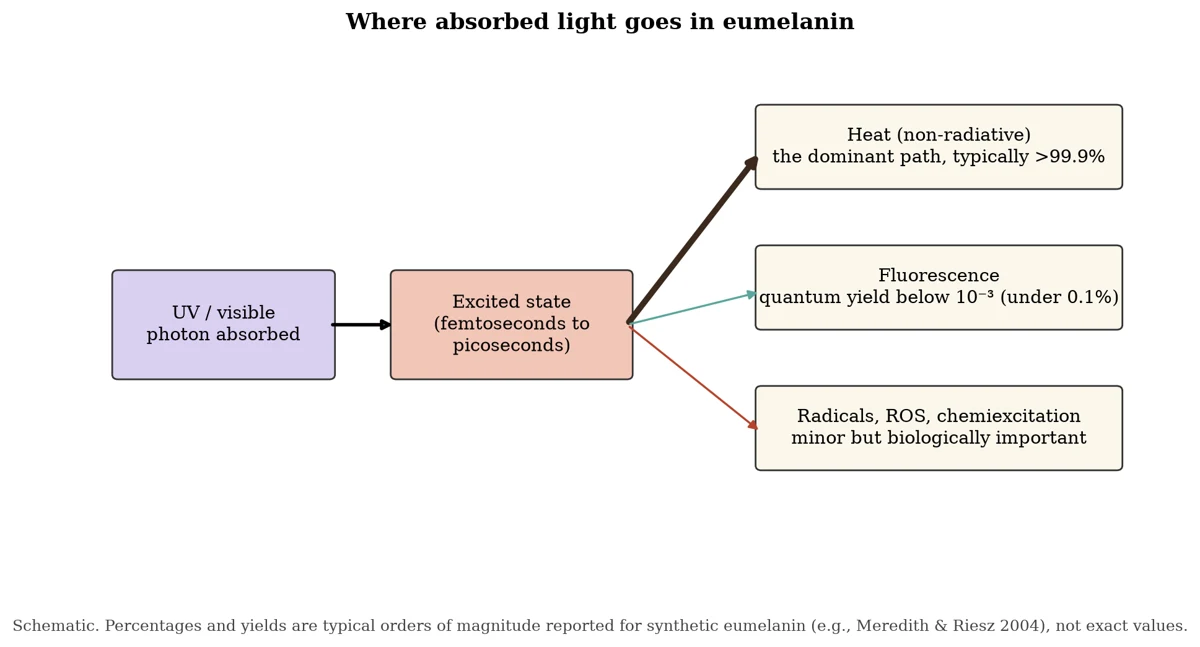
Absorbing light is only half of protection. A molecule that soaks up a UV photon holds a burst of energy that could break bonds or create reactive species. Melanin's answer is speed. Meredith and Jennifer Riesz reported in Photochemistry and Photobiology in 2004 that the radiative quantum yield of synthetic eumelanin, the share of absorbed light given back as fluorescence, is extremely low: eumelanin dissipates more than 99.9% of absorbed UV and visible light without emitting it, and the yield changes with the color of the exciting light. (Riesz, Sarna and Meredith later measured about 0.2% for synthetic pheomelanin, twice the eumelanin value.) The energy is lost very fast. Using femtosecond laser spectroscopy, Chris Grieco, Forrest Kohl and Bern Kohler at Ohio State reported in 2023 that light also creates radicals within the first moments after absorption, and Sarna, commenting on that work, described a new picture in which ultrafast deactivation comes from the rapid recombination of those promptly formed radicals. In Meredith and Sarna's account, this near-total non-radiative dissipation is what makes eumelanin such an effective photoprotectant.
The stable free radical
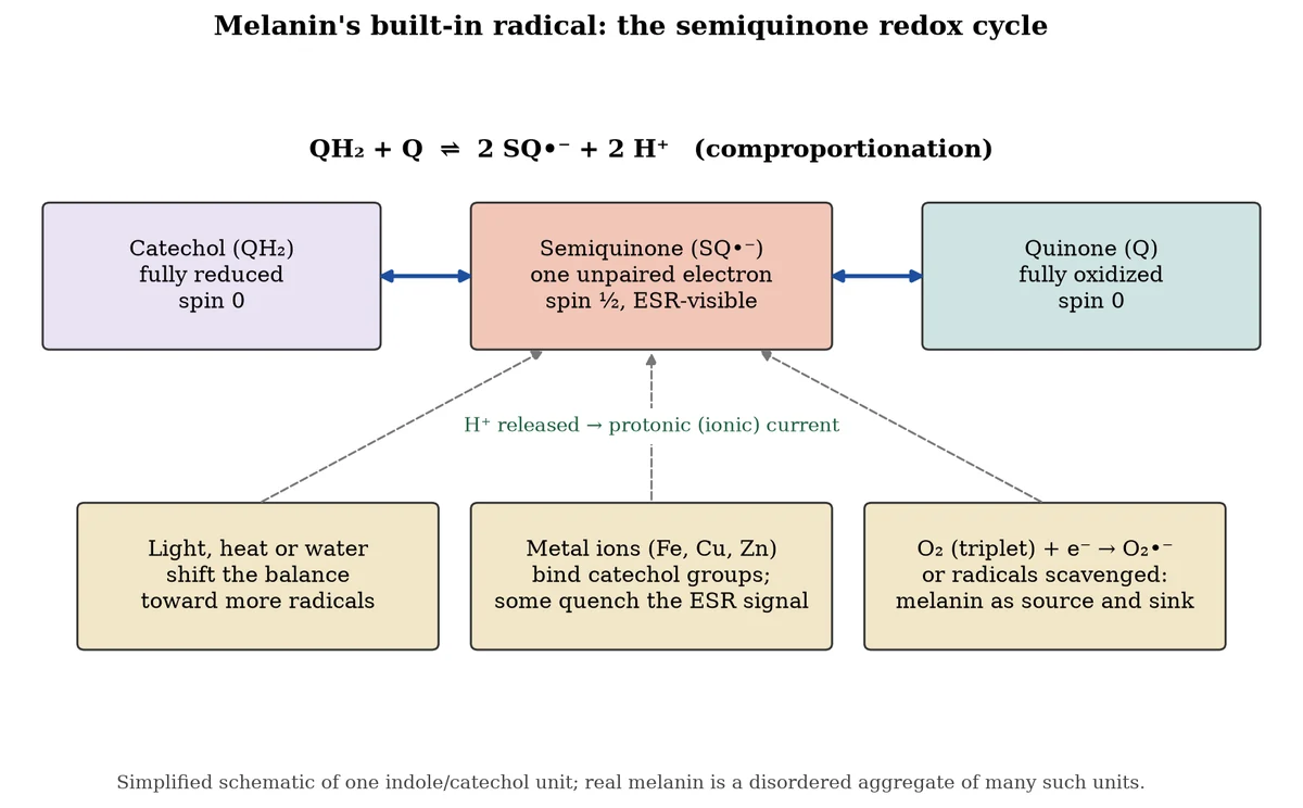
The quantum story proper begins with spin. In 1954, Barry Commoner, Jonathan Townsend and George Pake reported in Nature that freeze-dried biological tissues give electron spin resonance (ESR) signals, the microwave absorption of unpaired electrons sitting in a magnetic field. ESR works because an unpaired electron is a tiny magnet with spin ½: in a field it can point with or against, and microwaves of the right frequency flip it.
In 1960, Howard Mason, Dennis Ingram and B. Allen published "The free radical property of melanins" in Archives of Biochemistry and Biophysics. They found that melanins carry a strong, persistent ESR signal, unlike the fleeting radicals of most chemistry. Melanin, in other words, is a permanent home for unpaired electrons. Later work traced much of that signal to semiquinone units, partly reduced forms of the catechol and quinone groups in the polymer, and to carbon-centered radicals.
This ties directly to the oxygen spin article on this site. Oxygen in the air is a triplet with two unpaired electrons. Melanin's semiquinones carry one. When the two meet, an electron can pass from melanin to oxygen to make superoxide, or melanin can capture and neutralize other radicals. Both happen, as Pillar 2 explains.
A magnet for metals
Melanin's catechol and carboxyl groups bind metal ions tightly. In 1978, C. C. Felix, James Hyde, Tadeusz Sarna and R. C. Sealy reported ESR evidence in the Journal of the American Chemical Society for chelate complexes between melanin and metal ions, and showed that some paramagnetic metals change the pigment's own radical signal. Binding iron, copper, zinc and calcium is now regarded as one of melanin's core properties.
Where melanin lives in the body
- Skin and hair. Melanocytes make pigment in organelles called melanosomes and hand them to surrounding skin cells, where they cap the nucleus like a parasol against UV.
- The eye. The retinal pigment epithelium (RPE), the iris and the choroid hold melanin that absorbs stray light and, in the RPE, sits beside the most oxygen-rich, light-exposed tissue in the body. Ocular melanin's roles were reviewed by Dan-Ning Hu, John Simon and Sarna in Photochemistry and Photobiology in 2008.
- The inner ear. Melanocytes sit in the stria vascularis of the cochlea and in the dark cell zone of the vestibular organs. As Justine Renauld's group at Creighton University summarizes (2026), the stria vascularis pumps potassium into the cochlear fluid to create the endocochlear potential that hearing depends on, and defects in strial cells disrupt that ionic balance.
- The brain. Neuromelanin fills the substantia nigra, the "black substance" visible to the naked eye in a brain slice. Luigi Zecca, at Italy's National Research Council in Milan, and colleagues measured in the Journal of Neurochemistry in 1994 that neuromelanin holds large amounts of iron and other metals. In 2006, Makoto Sasaki and colleagues at Iwate Medical University reported in NeuroReport a 3-tesla MRI technique that shows neuromelanin-rich neurons in the substantia nigra and locus coeruleus as bright areas, and found the signal reduced in Parkinson's disease. The contrast is generally attributed to neuromelanin's complexes with paramagnetic iron. The neurons richest in neuromelanin are among the first lost in Parkinson's disease.
Pillar 2: Middle Ground: active and contested research
Middle Ground
Why so broad? Disorder versus excitons
For decades the smooth absorption was explained by analogy to semiconductors: a band structure, like silicon's, absorbing everything above a gap. Meredith's group proposed a different picture. In 2006, M. Linh Tran, Ben Powell and Paul Meredith argued in the Biophysical Journal that eumelanin is a chemically and structurally disordered mix of many slightly different small units, each absorbing in its own narrow window, and that the sum of all of them produces the broad curve. They called this the chemical disorder model.
In 2014, Chun-Teh Chen, Markus Buehler and colleagues at MIT reported in Nature Communications, from computer models, that the stacking geometry of eumelanin units, both orderly and disordered, creates excitonic effects that broaden absorption further. Today most researchers in the field treat disorder at several levels, chemical and geometric, as the main explanation. The older "true semiconductor band gap" picture has largely been set aside, though the exact balance among the contributing effects is still debated.
Semiconductor or wet ionic conductor?
In 1974, John McGinness, Peter Corry and Peter Proctor reported in Science that melanin pellets switch between low and high conductivity under voltage, like amorphous semiconductor devices. In 1960 the theoretical chemist H. Christopher Longuet-Higgins had published a short note in Archives of Biochemistry and Biophysics on the origin of melanin's free radical signal, and he is widely credited with first proposing that melanin might behave as a semiconductor. Together, the theory and the 1974 switching result made "melanin as a biological semiconductor" a lasting idea. As Mostert and colleagues put it in their 2012 paper, from the 1970s it was widely believed that melanins are naturally occurring amorphous organic semiconductors.
Then came the water problem. Melanin's conductivity rises steeply with hydration. In 2012, A. Bernardus Mostert, Meredith and colleagues reported in PNAS that much of melanin's conduction is ionic, carried by protons, rather than electronic. Using conductivity, muon spin relaxation and EPR measurements at different humidities, they concluded that melanin is an electronic-ionic hybrid conductor rather than an amorphous semiconductor. Their model links the two pictures: water drives the comproportionation reaction in Figure 3, in which a catechol and a quinone react to give two semiquinone radicals and release protons. More water means more radicals and more mobile protons. Mostert's team backed this with hydration-controlled ESR in The Journal of Physical Chemistry B (2013), which tracked the radical count rising as samples absorbed water, and in 2018 Margarita Sheliakina, Mostert and Meredith reported in Advanced Functional Materials a method for separating the ionic and electronic currents in the same sample.
In their 2021 review for materials scientists in Polymers, Mostert describes melanin as a hybrid ionic-electronic conductor, a property that has made it a candidate material for bioelectronics, where devices must talk to tissue in the language of ions. Pezzella and coworkers in Naples work on eumelanin thin films for the same reason. What fraction of the current is electronic, and how much any of this matters inside a living cell rather than a pressed pellet or film, remains open.
Shield and source: melanin and reactive oxygen
Melanin both quenches and makes reactive oxygen species. Its radicals can mop up other radicals, and its metal binding can lock away iron that would otherwise drive the Fenton reaction. But its semiquinones can also hand an electron to oxygen, especially under light, producing superoxide and hydrogen peroxide. In 2002, Małgorzata Różanowska, Sarna, Michael Boulton and colleagues reported in Investigative Ophthalmology & Visual Science that blue light makes melanosomes from the retinal pigment epithelium consume oxygen and produce superoxide and hydrogen peroxide, and that this photoreactivity rises with the donor's age, though it stayed well below that of the age pigment lipofuscin. Pheomelanin is generally reported to be more pro-oxidant than eumelanin. Which role dominates depends on the pigment type, its age, its metal load and the light it sees.
Chemiexcitation: the dark side of a dark pigment
In 2015, Sanjay Premi, Douglas Brash and colleagues at Yale reported in Science that melanin can cause DNA damage hours after UV exposure ends. In mouse and human melanocytes, UV triggers chemistry that excites melanin fragments into high-energy states, without any further light, and that energy passes to DNA to form the same cyclobutane pyrimidine dimers that sunlight makes directly. They called the process chemiexcitation. The finding is replicated in parts but its weight in human skin cancer is still under study.
Neuromelanin: guardian or culprit
Zecca's group reported in PNAS in 2008 that pigments related to neuromelanin build up across many brain regions with age and trap toxic metals. In their view, neuromelanin starts life as a protective sink for iron and stray dopamine-derived quinones. When neurons die and release it, the pigment, now loaded with iron, may feed inflammation. Whether neuromelanin protects or harms, or does each at different stages, is an open question in Parkinson's research.
Radiotrophic fungi
In 2000, Nelli Zhdanova and colleagues in Kyiv reported in Mycological Research that the walls of the damaged Chernobyl reactor's containment were colonized by many fungal species, a large share of them heavily melanized. In 2007, Ekaterina Dadachova, Arturo Casadevall and colleagues at the Albert Einstein College of Medicine reported in PLoS ONE that ionizing radiation changes the ESR signal of melanin and its electron transfer properties, giving irradiated melanin a fourfold greater capacity to reduce NADH. Melanized Wangiella dermatitidis and Cryptococcus neoformans exposed to radiation about 500 times background grew significantly faster than non-irradiated melanized cells or irradiated albino mutants, with more colonies, more biomass and threefold greater uptake of carbon-14-labeled acetate. In a 2008 review in Current Opinion in Microbiology, Dadachova and Casadevall proposed that such fungi might capture energy from radiation through melanin, a process they called "radiosynthesis." Radames Cordero later suggested in Environmental Microbiology (2017) that fungal melanin could help shield astronauts. The protective effect is well supported. Whether the fungi actually harvest usable metabolic energy from radiation has not been shown by direct energy accounting, and the researchers themselves present it as a hypothesis.
Pillar 3: Decentralized View: emerging and unestablished ideas
Decentralized View
Melanin as an energy transducer
The researchers in this pillar take melanin's physics as a starting point and go much further. Mexican ophthalmologist Arturo Solís Herrera has argued since the early 2000s that melanin splits water when it absorbs light, producing hydrogen, oxygen and high-energy electrons, so that human cells harvest light energy chemically, a process he calls human photosynthesis. He set out the claim in a 2015 paper in Integrative Cancer Science and Therapeutics titled "The unsuspected intrinsic property of melanin to transform light energy into chemical energy and the Warburg effect." Mainstream melanin researchers have not confirmed water splitting by melanin in living tissue, and Solís Herrera's papers appear mostly outside the core pigment-cell literature.
Dr. Jack Kruse: melanin, light and mitochondria
Dr. Jack Kruse, a neurosurgeon who writes about quantum biology, places melanin at the center of his account of how light powers life. In his 2017 post "Reality #15: Animal Photosynthesis," he writes, "We use hydrated proteins made from aromatic amino acids to separate water to make energy in human cells," and, of UV light and melanin, "This is why UV light and melanin are so helpful in repair defective mitochondria." He frames the pigment as an engine of light-driven energy: "How melanin works photosynthetically in us is one small example."
In Kruse's view, sunlight on the skin and eye drives melanin to dissociate water, release electrons, shift redox balance and tune how metals such as iron and copper are held, which in turn controls how mitochondria use oxygen. He links this with the leptin–melanocortin pathway, in which UV light triggers POMC and α-MSH to raise melanin production, and with his broader argument that a low-light modern life starves this circuit. In a January 2026 LinkedIn post announcing a blog on melanin evolution, he writes that emerging views in quantum biology suggest melanin dissociates water on absorbing light. That water-splitting claim is the same one Solís Herrera makes; the peer-reviewed work in Pillar 2 shows water driving radical and proton chemistry in melanin, not net water splitting.
Neuromelanin, spin and the quantum brain
Some writers in quantum biology propose that neuromelanin's stable radicals and bound iron give it quantum roles in the brain: as a spin-carrying antenna, an electron or proton conduit, or a link between light, magnetism and dopamine neurons. The substantia nigra does hold a paramagnetic, iron-rich pigment that MRI can image, and the pigment does carry stable spins. No published study we found shows neuromelanin performing a quantum function in signaling or cognition.
Radical pairs and magnetic sensitivity
The oxygen spin article described the radical-pair mechanism: two radicals born together whose spins, and therefore whose chemistry, can be nudged by a magnetic field. Melanin produces radicals and reacts with oxygen, so some researchers and writers have asked whether it could be magnetically sensitive in the same way. ESR itself proves melanin's spins respond to magnetic fields, but at strengths far above Earth's. A demonstrated radical-pair magnetic effect in melanin, at Earth-strength fields, was not found in the literature reviewed for this draft.
How the threads connect
The featured researchers draw these strands together in different ways. For Mason and Sarna, melanin is a stable radical and an efficient converter of light to heat. For Meredith and Mostert, it is a disordered, water-driven ionic-electronic material. For Dadachova and Casadevall, it is a shield that may let fungi use radiation. For Solís Herrera and Kruse, it is a light-to-chemical-energy transducer at the heart of human energy metabolism.
Summary table
| Claim | Label |
|---|---|
| Melanin forms by the tyrosinase (Raper–Mason) pathway as eumelanin, pheomelanin and neuromelanin | Centralized View |
| Broadband UV-visible absorption; light converted almost entirely to heat | Centralized View |
| Melanin carries stable free radicals detectable by ESR (Mason 1960) | Centralized View |
| Melanin binds metals; iron-laden neuromelanin is visible on MRI | Centralized View |
| Broad absorption explained by chemical and geometric disorder | Middle Ground |
| Hydration-dependent conduction is largely protonic, tied to comproportionation (Mostert) | Middle Ground |
| Melanin both quenches and generates ROS; chemiexcitation (Premi, Brash) | Middle Ground |
| Melanized fungi grow better under ionizing radiation (Dadachova) | Middle Ground |
| "Radiosynthesis": fungi harvest energy from radiation | Middle Ground Decentralized View |
| Melanin splits water to power human cells (Solís Herrera, Kruse) | Decentralized View |
| Neuromelanin quantum functions; Earth-field radical-pair effects in melanin | Decentralized View |
This article presents viewpoints, attributed to their sources. It is not medical advice.
Sources
- Raper, H. S. (1927). The tyrosinase-tyrosine reaction. Biochemical Journal 21, 89–96. https://doi.org/10.1042/bj0210089
- Commoner, B., Townsend, J. & Pake, G. E. (1954). Free radicals in biological materials. Nature 174, 689–691. https://doi.org/10.1038/174689a0
- Mason, H. S., Ingram, D. J. E. & Allen, B. (1960). The free radical property of melanins. Archives of Biochemistry and Biophysics 86, 225–230. https://doi.org/10.1016/0003-9861(60)90409-4
- Piattelli, M. & Nicolaus, R. A. (1961). The structure of melanins and melanogenesis—I. Tetrahedron 15, 66–75. https://doi.org/10.1016/0040-4020(61)80009-4
- McGinness, J., Corry, P. & Proctor, P. (1974). Amorphous semiconductor switching in melanins. Science 183, 853–855. https://doi.org/10.1126/science.183.4127.853
- Felix, C. C., Hyde, J. S., Sarna, T. & Sealy, R. C. (1978). Interactions of melanin with metal ions. Journal of the American Chemical Society 100, 3922–3926. https://doi.org/10.1021/ja00480a044
- Prota, G. (1988). Progress in the chemistry of melanins and related metabolites. Medicinal Research Reviews 8, 525–556. https://doi.org/10.1002/med.2610080405
- Zecca, L. et al. (1994). Iron and other metals in neuromelanin, substantia nigra, and putamen of human brain. Journal of Neurochemistry 62, 1097–1101. https://doi.org/10.1046/j.1471-4159.1994.62031097.x
- Zhdanova, N. N. et al. (2000). Fungi from Chernobyl: mycobiota of the inner regions of the containment structures of the damaged nuclear reactor. Mycological Research 104, 1421–1426. https://doi.org/10.1017/S0953756200002756
- Ito, S. & Wakamatsu, K. (2003). Quantitative analysis of eumelanin and pheomelanin in humans, mice, and other animals: a comparative review. Pigment Cell Research 16, 523–531. https://doi.org/10.1034/j.1600-0749.2003.00072.x
- Meredith, P. & Riesz, J. (2004). Radiative relaxation quantum yields for synthetic eumelanin. Photochemistry and Photobiology 79, 211–216. https://doi.org/10.1562/0031-8655(2004)079<0211:RCRQYF>2.0.CO;2
- Meredith, P. & Sarna, T. (2006). The physical and chemical properties of eumelanin. Pigment Cell Research 19, 572–594. https://doi.org/10.1111/j.1600-0749.2006.00345.x
- Tran, M. L., Powell, B. J. & Meredith, P. (2006). Chemical and structural disorder in eumelanins: a possible explanation for broadband absorbance. Biophysical Journal 90, 743–752. https://doi.org/10.1529/biophysj.105.069096
- Dadachova, E. et al. (2007). Ionizing radiation changes the electronic properties of melanin and enhances the growth of melanized fungi. PLoS ONE 2, e457. https://doi.org/10.1371/journal.pone.0000457
- Dadachova, E. & Casadevall, A. (2008). Ionizing radiation: how fungi cope, adapt, and exploit with the help of melanin. Current Opinion in Microbiology 11, 525–531. https://doi.org/10.1016/j.mib.2008.09.013
- Hu, D.-N., Simon, J. D. & Sarna, T. (2008). Role of ocular melanin in ophthalmic physiology and pathology. Photochemistry and Photobiology 84, 639–644. https://doi.org/10.1111/j.1751-1097.2008.00316.x
- Zecca, L. et al. (2008). New melanic pigments in the human brain that accumulate in aging and block environmental toxic metals. PNAS 105, 17567–17572. https://doi.org/10.1073/pnas.0808768105
- Simon, J. D. et al. (2009). Current challenges in understanding melanogenesis. Pigment Cell & Melanoma Research 22, 563–579. https://doi.org/10.1111/j.1755-148X.2009.00610.x
- Mostert, A. B., Powell, B. J., Pratt, F. L., Hanson, G. R., Sarna, T., Gentle, I. R. & Meredith, P. (2012). Role of semiconductivity and ion transport in the electrical conduction of melanin. PNAS 109, 8943–8947. https://doi.org/10.1073/pnas.1119948109
- Mostert, A. B. et al. (2013). Hydration-controlled X-band EPR spectroscopy. The Journal of Physical Chemistry B 117, 4965–4972. https://doi.org/10.1021/jp401615e
- d'Ischia, M. et al. (2013). Melanins and melanogenesis: methods, standards, protocols. Pigment Cell & Melanoma Research 26, 616–633. https://doi.org/10.1111/pcmr.12121
- Chen, C.-T. et al. (2014). Excitonic effects from geometric order and disorder explain broadband optical absorption in eumelanin. Nature Communications 5, 3859. https://doi.org/10.1038/ncomms4859
- Premi, S. et al. (2015). Chemiexcitation of melanin derivatives induces DNA photoproducts long after UV exposure. Science 347, 842–847. https://doi.org/10.1126/science.1256022
- Solís Herrera, A. (2015). The unsuspected intrinsic property of melanin to transform light energy into chemical energy and the Warburg effect. Integrative Cancer Science and Therapeutics 2(5). https://doi.org/10.15761/ICST.1000152
- Cordero, R. J. B. (2017). Melanin for space travel radioprotection. Environmental Microbiology 19, 2529–2532. https://doi.org/10.1111/1462-2920.13753
- Sheliakina, M., Mostert, A. B. & Meredith, P. (2018). Decoupling ionic and electronic currents in melanin. Advanced Functional Materials 28, 1805514. https://doi.org/10.1002/adfm.201805514
- Mostert, A. B. (2021). Melanin, the what, the why and the how: an introductory review for materials scientists interested in flexible and versatile polymers. Polymers 13, 1670. https://doi.org/10.3390/polym13101670
- Longuet-Higgins, H. C. (1960). On the origin of the free radical property of melanins. Archives of Biochemistry and Biophysics 86, 231–232. https://doi.org/10.1016/0003-9861(60)90410-0
- Różanowska, M. et al. (2002). Photoreactivity of aged human RPE melanosomes: a comparison with lipofuscin. Investigative Ophthalmology & Visual Science 43(7), 2088–2096. PMID 12091401
- Sasaki, M. et al. (2006). Neuromelanin magnetic resonance imaging of locus ceruleus and substantia nigra in Parkinson's disease. NeuroReport 17(11), 1215–1218. https://doi.org/10.1097/01.wnr.0000227984.84927.a7
- Riesz, J., Sarna, T. & Meredith, P. (2006). Radiative relaxation in synthetic pheomelanin. The Journal of Physical Chemistry B 110(28), 13985–13990. https://doi.org/10.1021/jp054869l
- Grieco, C., Kohl, F. R. & Kohler, B. (2023). Ultrafast radical photogeneration pathways in eumelanin. Photochemistry and Photobiology 99(2), 680–692. https://doi.org/10.1111/php.13731
- Sarna, T. (2023). Photodynamics of melanin radicals: contribution to photoprotection by melanin. Photochemistry and Photobiology 99(2), 866–868. https://doi.org/10.1111/php.13753
- Nayak, M. K. et al. (2026). Targeting the melanocytes of the inner ear: a comparison of different CreER lines. Developmental Biology 530, 224–233. https://doi.org/10.1016/j.ydbio.2025.12.009
- Kruse, J. (2017, Aug 11). Reality #15: Animal Photosynthesis. jackkruse.com/reality-15-animal-photosynthesis/
- Kruse, J. (2026, Jan). LinkedIn post announcing a melanin-evolution blog. linkedin.com/posts/drjackkruse_decentralized-medicine-92-melanin-evolution-activity-7421914877675520000-I-V2
All DOIs, volumes, issues, pages, years and author names above were checked against Crossref and PubMed records on Nov 3, 2026.
Not yet verified
- Direct quotes: the only direct quotes are Dr. Jack Kruse's three sentences from "Reality #15," checked word for word against the live page on Nov 3, 2026. Every other researcher's view is paraphrased. The January 2026 LinkedIn post is paraphrased from a search-index copy; the page itself was not opened, and its date comes from the index.
- Longuet-Higgins (1960): the note is real and cited, but it has no abstract online and the full text was not read, so his semiconductor proposal is given as the attribution commonly made in the literature, not confirmed from the paper itself.
- Mostert and water splitting: no Mostert paper reporting that melanin splits water was found. His verified work covers hydration-driven comproportionation and protonic conduction, and the article describes only that.
- Commoner 1954, Mason 1960, Zhdanova 2000, Felix 1978, Ito & Wakamatsu 2003, Zecca 1994/2008, Chen 2014, Premi 2015, Sheliakina 2018: bibliographic details were confirmed, but the descriptions rest on titles, abstracts and standard summaries, not full texts. Zhdanova's species counts are not given.
- First names not in records: Crossref and PubMed give only initials for B. Allen (Mason 1960), C. C. Felix and R. C. Sealy, so the article uses initials. "Dennis" Ingram (D. J. E. Ingram) and "Christopher" Longuet-Higgins are their known first names, not shown in the records.
- Raper–Mason pathway: the name is widely used, but no source is cited for it.
- Diagrams: all three figures are schematics made for this draft. The curves in Figure 1 are drawn, not measured. Figure 2's percentages are typical orders of magnitude.
Better signals, less noise.
One email a week: the best threads on light, Earth cycles and sovereignty, with context, evidence labels and links to the original sources.
Thanks! You're on the list.
Sorry, something went wrong. Please try again, or reach us on X at @primal_buddha.
No spam. Unsubscribe anytime.