 Primal Adapted
Light From Within: The Quantum Biophysics of Ultraweak Photon Emission (UPE)
Living tissue emits a steady trickle of photons from its oxygen chemistry, with mitochondria among the main sources. Here is what researchers have measured, what they debate, and the less established idea that this internal light does work inside the body.
Close the door of a perfectly dark room and wait. After half an hour your eyes have adapted as far as they can, and you see nothing. Yet a camera cooled to a hundred degrees below zero, pointed at your face, slowly builds an image of it in light. Masaki Kobayashi and his colleagues in Japan put it plainly in 2009: the human body glimmers, at an intensity about a thousand times below what the naked eye can detect. Every living thing tested so far does the same. Yeast, seedlings, a rat's brain, a cut leaf and the back of a human hand all give off a faint, steady trickle of photons that has nothing to do with heat glow or with the bright bioluminescence of fireflies.
Scientists call this ultraweak photon emission, or UPE. Many in the field also call it biophoton emission. Its existence is no longer in doubt. What it means is another matter. To most chemists it is the light given off when oxygen chemistry inside cells goes slightly off course, a by-product of metabolism that can be used as a sensor of oxidative stress. To a smaller group of physicists and neuroscientists it may be a signal: light that neurons could pass along their axons, that mitochondria could guide through their own networks, or that cells could use to sense each other. To a third group it is far more: the visible edge of a coherent field of light that organizes the body.
One idea from that third group gives this article its theme: that mitochondria use UPEs to "tan" the insides. The picture is of an internal light source: mitochondria making photons that then bathe nearby tissue from within, a kind of inner tanning, acting on the same light-absorbing molecules that sunlight acts on from outside. To be clear from the start, "tanning the insides" is a Decentralized View framing used informally in parts of the quantum-biology community. It is not an established biological process, and no study found for this article has measured it. What is established is narrower, and still remarkable. Cells do emit ultraweak light. Most of that light comes from reactive oxygen chemistry, much of it in or around mitochondria. Red and near-infrared light from outside the body can change how cells behave, a field called photobiomodulation. And one of the main molecules thought to absorb that light is cytochrome c oxidase, the final enzyme of the mitochondrial respiratory chain.
This article follows the light through three labels used across the site:
- Centralized View covers what is well established: how UPE is measured, the chemistry that makes it, and how external red and near-infrared light acts on cells through cytochrome c oxidase.
- Middle Ground covers real, published research that is still contested: neural UPE and the idea of optical channels in nerves, mitochondria as waveguides, claims about coherence, UPE as a diagnostic, the long dispute over Gurwitsch's mitogenetic rays, and quantum ideas about microtubules.
- Decentralized View covers ideas that have not been established: Fritz-Albert Popp's coherent biophoton field, DNA as a store of light, the mitochondria-as-inner-light-source or "inner tanning" picture, Dr. Jack Kruse's account of biophotons and mitochondria, and related ideas about melanin and water.
Where a finding spans two labels, the text says which part belongs where. Where the research has not shown a link, the text says so plainly. This article connects to three others on this site: the oxygen spin article, on how oxygen's unpaired electrons shape mitochondrial chemistry; the earthing article, on charge, water and the body; and The Dark Battery, on melanin's stable radicals.
Pillar 1: Centralized View: the established science
Centralized View
The Centralized View of ultraweak photon emission rests on three things: instruments that can count single photons in the dark, a well-mapped chemistry of excited molecules, and a separate but related field showing that cells respond to red and near-infrared light. Each is described here as researchers in those fields describe it.
A century of looking for faint light
The story begins in the 1920s with the Russian biologist Alexander Gurwitsch. Working with onion roots, he reported that the tip of one growing root could speed up cell division in a second root nearby, even through quartz, which passes ultraviolet light, but not through ordinary glass, which blocks it. He concluded that dividing cells give off weak ultraviolet light that triggers division in their neighbors, and he called it mitogenetic radiation. In 1933 he reported in Nature that nerve tissue gave off the same kind of radiation. The claim set off a wave of work in the 1920s and 1930s, and a wave of failed replications that is told in the Middle Ground below.
Detecting that light directly had to wait for better instruments. In 1954 the Italian physicists L. Colli and U. Facchini used a photomultiplier tube to record light from germinating plant seedlings, a result they reported in Il Nuovo Cimento. In 1974 Terence Quickenden and Shane Que Hee in Australia reported weak luminescence from growing yeast and, two years later, measured its spectrum. Their work tied the emission to growth and oxidative metabolism rather than to a special mitogenetic ray.
In 1980 Enrique Cadenas, Alberto Boveris and Britton Chance at the University of Pennsylvania showed that particles made from beef heart mitochondria glow faintly when fed organic hydroperoxides. Blocking the respiratory chain with antimycin or rotenone raised the light by 50–60 percent. Adding cytochrome c raised it twelvefold. Quenchers of singlet oxygen changed it, and the authors concluded that singlet oxygen was mainly responsible for the light in that system. This put mitochondria and oxygen chemistry at the center of the story early on.
In Japan, Humio Inaba's group built what he called super-high-sensitivity photon counting systems, able to register count rates as low as one photoelectron per second to one per minute, and used them on blood and organ samples from rats. Inaba's former colleague Masaki Kobayashi then made images. In 1999 his team published the first two-dimensional images of spontaneous photon emission from a living rat's brain. In 2009 Kobayashi, Daisuke Kikuchi and Hitoshi Okamura imaged the light from human volunteers over the course of a day and found that it rose and fell in a daily rhythm.
Through the 2000s and 2010s, Pavel Pospíšil, Ankush Prasad and Michal Cifra in the Czech Republic did much of the careful work of tying the light to specific molecules, and Roeland van Wijk in the Netherlands spent decades measuring human emission. In 2014 Cifra and Pospíšil published a review that is now the standard reference for what UPE is and is not. Their definition is simple. Ultraweak photon emission is chemiluminescence from biological systems, produced when electronically excited molecules form during normal oxidative metabolism or during oxidative stress.
How you count light that faint
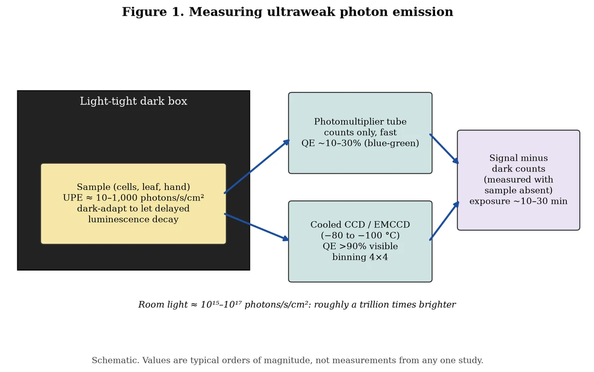
The numbers explain why it took so long. Cifra and Pospíšil put the intensity of UPE at several units to several hundred photons per second per square centimeter during normal oxidative metabolism, and several hundred to several thousand during oxidative stress. In 2025 Vahid Salari, Christoph Simon, Daniel Oblak and colleagues at the University of Calgary used the round figure of 10 to 1,000 photons per square centimeter per second. A typical room light delivers something like 10^15 to 10^17 photons per square centimeter per second. Biophotons are roughly a trillion times fainter.
Two kinds of detector do the work.
- Photomultiplier tubes (PMTs). A photon hits a photocathode and knocks out an electron. A chain of electrodes multiplies that one electron into a pulse of millions, which electronics count. PMTs are fast and sensitive but give no image, only a count over an area. Their quantum efficiency, the fraction of incoming photons that produce a count, is typically in the tens of percent in the blue and green and falls off in the red.
- Cooled CCD and EMCCD cameras. These are chip cameras cooled far below room temperature to suppress the thermal electrons that would otherwise swamp the signal. An electron-multiplying CCD (EMCCD) adds a gain stage on the chip so that single photons stand out above the readout noise. Salari and colleagues reported using EMCCD and CCD cameras with quantum efficiencies above 90 percent for visible light.
The practical limits are the same for both. Every detector produces dark counts, false signals that arise even in total darkness, from heat and from stray radioactivity. A measurement means nothing unless the dark count has been measured just before and after, with the sample absent. Samples must sit in total darkness long enough for delayed luminescence, the slowly fading glow left over from any earlier exposure to light, to die away. Imaging usually needs long exposures and binning, which combines neighboring pixels into one larger pixel to collect enough photons. In 2013 Prasad and Pospíšil reported that with 4 × 4 binning, a good image of spontaneous emission took about 30 minutes for yeast, 20 minutes for a human hand and 10 minutes for an Arabidopsis plant.
The spectrum and the difference between spontaneous and delayed light
Cifra and Pospíšil describe UPE as spanning the near ultraviolet, the visible and the near infrared, roughly 350 to 1,300 nanometers. Salari's group gives 200 to 1,000 nanometers as the range in which UPE has been observed. The exact limits depend on the detector, since few instruments are sensitive across the whole range, and on the sample.
Researchers keep two phenomena apart.
- Spontaneous UPE is the light a sample gives off in steady darkness, without being lit first. It reflects ongoing chemistry.
- Delayed luminescence is the light that continues for seconds to minutes after a sample has been illuminated. It decays over time, and its decay curve carries information about how the sample stores and releases excitation energy. Popp's school used delayed luminescence extensively, as the Middle Ground and Decentralized View sections describe.
Both are real and measurable. They are not the same thing, and confusing them is one of the more common errors in popular writing about biophotons.
Photon statistics: what Poisson means
A photon counter records how many photons arrive in each short time window. If you repeat that many times, you get a distribution of counts. For light from a laser in a so-called coherent state, the counts follow a Poisson distribution, in which the variance equals the mean. For ordinary thermal light measured over windows much longer than its coherence time, the counts also come out very close to Poisson. For chaotic light measured over very short windows, the counts are more spread out (variance greater than the mean). Certain non-classical states of light, called squeezed or sub-Poissonian states, are narrower.
This matters because Popp and others have argued that UPE counts fit Poisson statistics and therefore show coherence. In their 2015 critical review in the Journal of Luminescence, Cifra, Christian Brouder, Michaela Nerudová and Ondřej Kučera went through the theory and the experiments. Their conclusion, in the published abstract, is that UPE itself is experimentally well established, but that "no reliable evidence for the coherence or nonclassicality of UPE has actually been achieved up to now." Their main point is that a Poisson count distribution by itself does not tell coherent light apart from many ordinary sources. The coherence debate is covered in the Middle Ground below.
Where the light comes from: excited molecules
Most molecules sit in their lowest energy state. To emit visible light, a molecule must first be pushed into an electronically excited state and then fall back. In fluorescence, the push comes from absorbing light. In chemiluminescence, it comes from a chemical reaction energetic enough to leave a product molecule excited. Ultraweak photon emission is chemiluminescence, and the energetic reactions are those of reactive oxygen species (ROS).
Pospíšil, Prasad and Marek Rác reviewed the chemistry in 2014. Their account runs in steps.
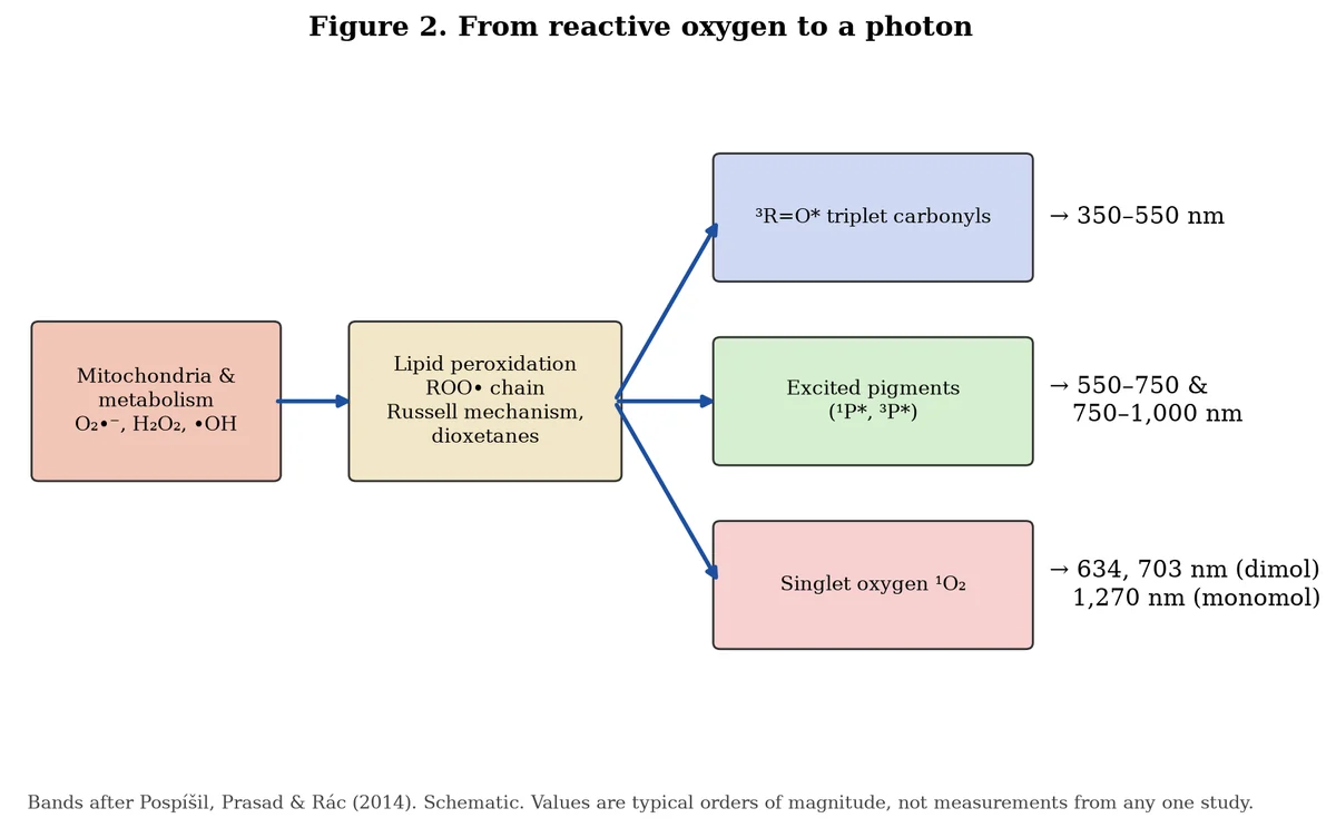
1. Initiation. Superoxide (O2•−), hydrogen peroxide and the hydroxyl radical (•OH) form during normal metabolism and in greater amounts during stress. Superoxide and especially hydroxyl radicals pull hydrogen atoms off lipids, proteins and nucleic acids. Singlet oxygen can also attack double bonds directly.
2. Propagation. In membranes, this sets off lipid peroxidation, a chain reaction in which lipid radicals react with oxygen to form peroxyl radicals (ROO•), which attack the next lipid.
3. Excitation. When two peroxyl radicals meet, they can combine through a short-lived four-oxygen intermediate (the Russell mechanism) that breaks apart into a ketone in its triplet excited state, an alcohol and, in some cases, singlet oxygen. Unstable four-membered rings called dioxetanes, formed when singlet oxygen adds to a double bond, also break down into triplet excited carbonyls.
4. Emission. Each excited species falls back to its ground state and may release a photon, or hand its energy to a nearby pigment that then emits.
The colors map onto the emitters. Pospíšil and colleagues assign triplet excited carbonyls to the near ultraviolet and blue-green (about 350–550 nm), singlet and triplet excited pigments to the green-red (550–750 nm) and red to near-infrared (750–1,000 nm), and singlet oxygen to the red at 634 and 703 nanometers and the near infrared at 1,270 nanometers.
Singlet oxygen's two lines need a word. The 1,270 nm line comes from a single singlet oxygen molecule returning to the ordinary triplet state. The 634 and 703 nm lines come from two singlet oxygen molecules colliding and releasing their energy as one photon, a process called dimol emission. The oxygen spin article on this site explains why oxygen's ground state is a triplet, why the singlet form is so reactive, and why the change between them is "spin-forbidden" and therefore slow. That spin rule is part of why excited oxygen and excited carbonyls live long enough to emit at all.
In 2018 Prasad, Anastasiia Balukova and Pospíšil tested this chain in pig skin. Hydroxyl radicals generated by the Fenton reaction started lipid peroxidation, and the skin gave off UPE. A singlet oxygen scavenger reduced the emission, showing a singlet oxygen share. A filter passing only 340–540 nm showed a substantial share from triplet carbonyls. Their earlier imaging work on human skin had pointed the same way.
Mitochondria as a main source
Mitochondria are the cell's main consumer of oxygen and, in most cells, one of its main sources of ROS. Michael Murphy's 2009 review in the Biochemical Journal describes how electrons leak from complexes I and III of the respiratory chain to oxygen, forming superoxide, and how the rate depends on the membrane potential, the ratio of NADH to NAD+ and the local oxygen level. Helmut Sies and Dean Jones's 2020 review in Nature Reviews Molecular Cell Biology describes how hydrogen peroxide made this way also acts as a signal, not only as damage.
Several lines of evidence tie UPE to mitochondria. Cadenas, Boveris and Chance's 1980 work showed that mitochondrial membranes glow under oxidative conditions. Kobayashi's 1999 rat brain study found that removing glucose suppressed emission from brain slices, that high potassium first enhanced and then depressed it, and that rotenone, which blocks complex I, increased it. The authors took this as a sign of electron leakage from the respiratory chain and concluded that the light from brain slices originates in mitochondrial energy metabolism through the production of reactive oxygen. In Tang and Dai's 2014 brain slice work, described below, sodium azide, a cytochrome c oxidase inhibitor, combined with oxygen and glucose deprivation, blocked glutamate-induced emission.
Inside the Centralized View, then, mitochondria are a main source of UPE because they are a main source of the ROS chemistry that makes excited molecules. This is not the same as saying mitochondria make light on purpose, or that the light does anything once made. Those questions belong to the other two pillars.
Light as an input: photobiomodulation
The second established thread runs the other way: light going into cells rather than coming out.
Since the late 1960s, researchers have reported that low-power red and near-infrared light can speed wound healing, reduce pain and inflammation and protect stressed cells. The field was first called low-level laser therapy and is now called photobiomodulation, or PBM. The Russian biophysicist Tiina Karu did much of the early mechanistic work. In a 1999 review she proposed cytochrome c oxidase as a likely photoacceptor for monochromatic red to near-infrared light and laid out four primary mechanisms: changes in the redox state of respiratory chain components after their electronic states are excited, the generation of singlet oxygen, local transient heating of the absorbing molecules, and increased superoxide production with more hydrogen peroxide downstream. She described a secondary cascade of changes in pH, calcium, cyclic AMP and ATP that carries the signal through the cell.
Cytochrome c oxidase is a natural candidate. It holds two copper centers and two heme groups, and these absorb in the red and near infrared, the so-called optical window where tissue absorbs least and light penetrates deepest. Karu's action spectra, plots of a biological effect against wavelength, show peaks that line up with the enzyme's absorption bands. In a 2005 HeLa cell study with Lyudmila Pyatibrat and Natalia Afanasyeva, the increase in cell attachment after irradiation had maxima at 619, 657, 675, 740, 760 and 820 nanometers, which the authors attributed to cytochrome c oxidase.
Margaret Wong-Riley and colleagues tested the idea directly in 2005. They treated cultured neurons with 670 nm LED light after poisoning cytochrome c oxidase with potassium cyanide. The light partly restored enzyme activity and cut neuronal death from 83.6 to 43.5 percent at one cyanide dose, but the protection faded as the dose rose, consistent with cytochrome c oxidase being the target.
Nitric oxide and the redox signal
Nitric oxide binds to cytochrome c oxidase and competes with oxygen, slowing respiration. Light can dissociate nitric oxide from heme proteins, a fact long known from photochemistry. Michael Hamblin of the Wellman Center at Massachusetts General Hospital summarized the leading view in a 2018 review in Photochemistry and Photobiology: the primary site of light absorption in mammalian cells has been identified as the mitochondria and specifically cytochrome c oxidase, and it is hypothesized that inhibitory nitric oxide can be dissociated from the enzyme, restoring electron transport and raising the mitochondrial membrane potential. Karu, Pyatibrat and Afanasyeva's 2005 study found that nitric oxide donors added before irradiation shifted the action spectrum and removed the light-induced effect, which they read as support for a nitric oxide role.
Hamblin's 2017 review in AIMS Biophysics describes the downstream effects as a brief burst of ROS, more ATP, more nitric oxide and changes in calcium, followed by changes in transcription factors. It also notes a strongly biphasic dose response, in which low doses stimulate and high doses inhibit, and a striking difference between healthy and stressed cells. PBM can raise ROS in normal cells but lower them in oxidatively stressed cells. Joseph Tafur and Paul Mills's 2008 review placed PBM inside the wider field of redox signaling, the same field that treats hydrogen peroxide as a messenger.
Cytochrome c oxidase is not the only target. Hamblin also names light-sensitive ion channels and opsins. In a 2014 review, Salvatore Passarella and Karu described a range of molecules in mitochondria and elsewhere in the cell as sensitive to red and near-infrared light.
Other internal absorbers: melanin and water
Two more absorbers matter for the "inner tanning" question.
Melanin. Eumelanin absorbs across the whole ultraviolet and visible spectrum and turns almost all of that energy into heat. In 2004 Paul Meredith and Jennifer Riesz measured its radiative quantum yield, the fraction of absorbed photons that come back out as light, and found it extremely low. Melanin is found not only in skin and the eye but also as neuromelanin in parts of the brain such as the substantia nigra, where Luigi Zecca and colleagues have measured high levels of bound iron and other metals. The Dark Battery covers melanin's stable radicals and electrical behavior in detail.
Melanin also links back to chemiluminescence. In 2015 Sanjay Premi, Douglas Brash and colleagues reported in Science that melanin derivatives in skin cells can become chemically excited hours after ultraviolet exposure ends. Superoxide and nitric oxide react to form peroxynitrite, which attacks melanin fragments and leaves them in excited triplet states. Those excited states then pass their energy to DNA, creating the same kind of DNA damage that sunlight causes directly, in the dark. The authors called this chemiexcitation. It is a published case of excited states made by ROS inside the body, in the dark, acting on a nearby molecule. That is the Centralized View core of a link between melanin and internally generated excited states. It does not show that melanin captures UPE, and the Decentralized View section covers that larger claim.
Water. Water absorbs strongly in the near infrared and beyond, especially above about 1,000 nanometers and very strongly near 1,450 nanometers. In the red and the first part of the near infrared it is nearly transparent, which is why those wavelengths reach deepest into tissue. The Middle Ground covers claims that light changes the structure of water at surfaces.
Pillar 2: Middle Ground: active and contested research
Middle Ground
The Middle Ground holds published, peer-reviewed research that is still debated: results that have not been widely reproduced, models that have not been tested in living animals, and interpretations that other researchers dispute.
The mitogenetic ray dispute
Gurwitsch's mitogenetic radiation is the cautionary tale of the field. In the late 1920s and early 1930s, hundreds of papers reported effects of mitogenetic rays on yeast, bacteria and tissues, and many others reported nothing. In 1931 Alexander Hollaender and Eugene Schoeffel reviewed the state of the field in The Quarterly Review of Biology, and in 1935 Hollaender and Walter Claus described their own attempts in the Journal of the Optical Society of America. Their work, along with other failures of the period, is widely cited as the reason interest faded outside the Soviet Union. The detectors of the time could not reliably record light that faint, and the biological assays used to detect it were hard to control.
In 1988 Gurwitsch's daughter, Anna Gurwitsch, published a historical review of the problem in Experientia, in the same issue as Inaba's description of modern photon counting. Her account presents the later direct photon measurements as support for the core claim that living cells emit weak ultraviolet light. Quickenden and Que Hee framed their 1974 yeast result as bearing on the existence of mitogenetic radiation without confirming that the light triggers division. That last step, that light from one cell causes another to divide, is still not established by modern, controlled experiments.
Neural UPE: glutamate, brain slices and spectra
In Wuhan, Jiapei Dai's group at South-Central University for Nationalities has published a series of studies on light in nerve tissue.
In 2010 Yan Sun, Chao Wang and Dai developed a technique they called in situ biophoton autography, which records photons by their effect on a silver-based photographic emulsion. In rat spinal nerve roots in vitro, they found that shining light on one end of a root increased photon activity at the other end. A nerve-blocking anesthetic (procaine) and metabolic inhibitors significantly reduced the effect. The authors concluded that light stimulation can generate biophotons that conduct along neural fibers, probably as neural communication signals.
In 2014 Rendong Tang and Dai imaged mouse brain slices. Long exposure to glutamate, the main excitatory neurotransmitter, gradually raised photon activity, peaking after about 90 minutes and lasting more than 200 minutes. Oxygen and glucose deprivation combined with sodium azide blocked the effect. Blocking action potentials with tetrodotoxin, applying procaine or removing calcium blocked it only partly. The authors reported that activity in the corpus callosum and thalamus appeared to travel along neural pathways.
In 2016, in PNAS, Zhuo Wang, Niting Wang, Zehua Li, Fangyan Xiao and Dai compared glutamate-induced emission in brain slices from bullfrogs, mice, chickens, pigs, monkeys and humans. They reported a spectral redshift along that sequence, reaching about 865 nm in human brain tissue, and proposed it may be a key biophysical basis for human intelligence. A comment published in PNAS the same year questioned that interpretation. The measurement has not been widely repeated outside Dai's group.
These studies are real and published. Their limits are that most come from one laboratory, the photographic method is indirect, and slices are not living brains. A rise in light after glutamate is also what the Centralized View would predict, since excitotoxic glutamate raises mitochondrial ROS. The open question is whether the light carries information, not whether it appears.
Axons as optical fibers
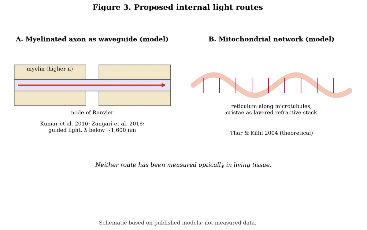
Can nerves guide light? In 2016 Sourabh Kumar, Kristine Boone, Jack Tuszyński, Paul Barclay and Christoph Simon, at the Universities of Calgary and Alberta, published "Possible existence of optical communication channels in the brain" in Scientific Reports. Using detailed optical models, they showed that myelinated axons could act as photonic waveguides even with realistic imperfections, because myelin has a higher refractive index than the surrounding fluid. They stated plainly that the work is theoretical, proposed experiments in vivo and in vitro to test it, and discussed whether photons could carry quantum entanglement in the brain.
In 2018 Andrea Zangari, Davide Micheli, Roberta Galeazzi and Antonio Tozzi modeled the node of Ranvier, the bare gap between segments of myelin, as an array of nanoscale antennas driven by ion currents. Their model suggested that wavelengths below about 1,600 nm would be most likely to travel through myelinated segments. In 2021 the same group, with additional colleagues, reported detecting photons at the node of Ranvier during electrically induced nerve activity using a silver photoreduction technique, and concluded that photonic radiation accompanies the action potential at the node.
Kumar's paper is a model. Zangari's 2018 paper is a model. Zangari's 2021 study and Sun's 2010 study are experiments using silver-grain methods, which can respond to chemistry as well as light. Direct optical measurement of light traveling along a living myelinated axon, with spectral and timing data, has not yet been reported.
Mitochondria as waveguides
In 2004 Roland Thar and Michael Kühl, at the University of Copenhagen's Marine Biological Laboratory, asked in the Journal of Theoretical Biology whether light could travel inside mitochondria. Their starting point was that mitochondria are the main source of ultraweak chemiluminescence from ROS. In vertebrate cells, mitochondria are often long filaments laid along microtubules, forming a connected network, the mitochondrial reticulum. Both mitochondria and microtubules have a higher refractive index than the surrounding cytoplasm, so in their model the network can act as an optical waveguide. They modeled the inner folds of the mitochondrion as a stack of layers with alternating refractive indices and showed that, if stimulated light emission could happen inside, such a structure could in principle support laser-like action, as in a distributed-feedback laser. They raised the question of whether some long-range interaction between mitochondria might be carried by light, without claiming to have shown it.
The paper is a theoretical model. No experiment found for this article has measured light traveling along a mitochondrial network in a living cell, or stimulated emission from mitochondria.
Cells that seem to see
Guenter Albrecht-Buehler, a cell biologist at Northwestern University, reported a series of experiments suggesting that cells can sense light from other cells. In 1992 in PNAS, under the title "Rudimentary form of cellular 'vision'," he reported that cells could detect the orientation of other cells across a thin glass film. A thin metal coating blocked the effect, but a silicone coating did not, which he took to mean the signal was electromagnetic radiation in the red to infrared range. In 2005, again in PNAS, he reported that aggregating 3T3 cells seemed to detect each other over many cell diameters and that this range grew with the cells' near-infrared light scattering. He concluded that near-infrared scattering may mediate a long-range attraction between cells.
These results come from one laboratory and have not been widely repeated. They are often cited as support for light-based cell communication, but their mechanism is unknown.
Photon statistics and coherence
Popp argued from the 1970s on that biophotons come from a coherent field, like laser light, rather than from random chemical events. His group's main evidence was photocount statistics close to Poisson and delayed luminescence that decays along a hyperbola rather than an exponential curve.
Cifra and colleagues' 2015 critical review is the main counterweight. They sorted the experimental literature into work with solid, conventional interpretations and work with unconventional interpretations. They pointed out that thermal and chaotic light can also yield near-Poisson counts at the time resolution used, that hyperbolic decay has ordinary explanations, and that a real test of coherence would require measuring coherence time and length directly or measuring photon correlations. They concluded that UPE is well established but that reliable evidence for coherence or nonclassical light from biological systems had not been achieved. As of this draft, no later study found has reported a direct measurement of coherence in biophotons that overturns that conclusion.
UPE as a diagnostic
The diagnostic use of UPE follows directly from the Centralized View chemistry: more oxidative stress means more excited molecules, which means more light.
- In 2004 Motohiro Takeda, Kobayashi and colleagues at Tohoku University imaged tumors transplanted into mice. Emission correlated with tumor size at one week (correlation coefficient 0.73) and, compared with tissue slides, seemed to reflect how much of the tumor was viable. They proposed UPE imaging as an optical biopsy.
- In 2009 Kobayashi's team found a daily rhythm in human emission and suggested it might track changes in energy metabolism.
- In 2025 Salari, Simon, Oblak and colleagues imaged live versus dead mice and found significantly more emission from living animals. In plants, higher temperatures and injuries both increased emission, and chemical treatments changed it. A local anesthetic, benzocaine, applied to a wound produced the highest emission of the compounds tested. They proposed UPE imaging as a label-free way to image vitality and plant stress.
- In 2010 Tafur, Eduard van Wijk, Roeland van Wijk and Mills argued in Photomedicine and Laser Surgery that biophoton detection, as a non-invasive readout of tissue redox state, might help predict who responds to photobiomodulation.
These are real measurements. Their limit is that UPE is a general readout of oxidative chemistry. It rises with temperature, injury, oxygen and many disease states, so on its own it cannot say which process is responsible. No UPE test has been approved for clinical diagnosis.
Biophotons and vision
In the retina, rods occasionally fire in total darkness, producing "dark noise" that looks exactly like a real photon hit. István Bókkon, Salari, Tuszyński and István Antal argued in 2010 that biophoton intensity inside cells may be much higher than measured outside, because most photons are absorbed before they escape. On that basis they proposed that biophotons in visual cortex neurons might be numerous enough to form internal images during perception and imagery. Salari, Tuszyński, Bókkon and colleagues had also discussed in 2011 how photonic interactions might relate to the electrical activity of neurons. In 2016, Salari, Felix Scholkmann, Bókkon, Farhad Shahbazi and Tuszyński argued in PLOS ONE that thermal activation explains only part of retinal dark noise and that internal photons from UPE are a more likely cause.
In 2012 Blake Dotta, Kevin Saroka and Michael Persinger at Laurentian University reported in Neuroscience Letters that volunteers imagining light in a dark room gave off more photons from the head, and that this tracked changes in EEG power. They presented this as support for Bókkon's hypothesis. The study was small, and the result has not been widely repeated by other groups.
Microtubules and quantum consciousness
Stuart Hameroff and Roger Penrose's Orch OR theory holds that consciousness arises from orchestrated quantum processes in microtubules inside neurons. Their 2014 review in Physics of Life Reviews updated the theory and answered critics. Some biophoton researchers, including Tuszyński, have discussed whether light in neurons could play a part in such processes, and Kumar and colleagues raised the question of entanglement carried by photons.
The best-known objection is Max Tegmark's 2000 paper in Physical Review E. He calculated that quantum coherence in the brain would decay within about 10^-13 to 10^-20 seconds, far shorter than the 10^-3 to 10^-1 seconds over which neurons work, and concluded that the brain should be treated as a classical system. Hameroff and colleagues have disputed his assumptions. The debate is open, and no experiment has shown that biophotons take part in it.
Light and water at surfaces
Gerald Pollack's group reported in 2006 that a zone of water next to water-loving surfaces pushes out suspended particles over long distances, and Pollack has argued that light, especially infrared, builds this "exclusion zone." The earthing article on this site covers the debate over its cause. Separately, Andrei Sommer and colleagues reported in 2011 in The Journal of Physical Chemistry Letters, under the title "Breathing volume into interfacial water with laser light," on effects of laser light on thin layers of water at surfaces, and Sommer has proposed that changes in interfacial water could be part of how photobiomodulation works. Both lines are published. Neither is part of the standard account of photobiomodulation, and neither has shown that biophotons from inside the body act on water.
Pillar 3: Decentralized View: ideas not yet established
Decentralized View
The Decentralized View holds ideas that go well beyond what has been measured. They are presented here as their proponents present them, with a neutral note after each on what would test them.
Fritz-Albert Popp: the coherent biophoton field
Fritz-Albert Popp, a German biophysicist who worked at the University of Marburg and later founded the International Institute of Biophysics in Neuss, is the figure most closely tied to the word "biophoton." From the 1970s until his death in 2018, he argued that the faint light of living cells is not a waste product of chemistry but the visible trace of a coherent electromagnetic field inside the organism. In his view, this field coordinates biochemical reactions, synchronizes cell division and carries information within and between cells. He pointed to photocount statistics close to Poisson and to the hyperbolic decay of delayed luminescence as evidence that the field is coherent, like laser light, rather than chaotic. In his 1984 paper in Cell Biophysics with Werner Nagl, K. H. Li, Walter Scholz, O. Weingärtner and R. Wolf, he set out the case for biophoton emission as a general property of living systems.
Popp described health as a state of high coherence and disease as a loss of it. He suggested that cancer cells lose the ability to keep their light ordered, and that measuring biophotons could become a tool for judging food quality and health.
Testing note: Coherence can be measured directly, by recording the coherence time and length of the light or by second-order photon correlation measurements that tell coherent, thermal and nonclassical light apart. Cifra and colleagues' 2015 review described what such tests would need. A direct measurement of this kind on UPE, repeated by independent groups, would test Popp's central claim.
DNA as a photon store
Popp also proposed that DNA is a main source and store of biophotons. In his model, DNA's tightly stacked bases form a structure that can hold excitation energy, act something like a cavity for light and release it in an ordered way. In 1981 Martin Rattemeyer, Popp and Werner Nagl reported in Naturwissenschaften evidence of photon emission from DNA in living systems, based on experiments with ethidium bromide, a dye that slips between DNA's bases and changes its structure, which they linked to changes in emission.
Testing note: If DNA stores and releases biophotons, then changes in DNA structure, density or packing in living cells should change emission in a predictable way, with emitted light at wavelengths that match DNA-bound excited states. Spectral measurements with DNA structure controlled independently of ROS levels would separate a DNA source from ordinary ROS chemistry.
Mitochondria as an internal light source: the "inner tanning" picture
This is the idea in the title. It puts several Centralized View and Middle Ground findings in a row and asks whether they connect.
1. Mitochondria are a main source of the ROS chemistry that makes UPE.
2. Mitochondria, through cytochrome c oxidase, are the main absorbers of red and near-infrared light in photobiomodulation.
3. Some UPE falls in the same red and near-infrared range.
4. Thar and Kühl modeled mitochondrial networks as possible waveguides.
5. Bókkon, Salari and colleagues argued that light intensity inside cells may be much higher than what escapes.
In the Decentralized View, these steps are joined. Mitochondria make light, the light travels through the cell and nearby tissue, and it is absorbed by cytochrome c oxidase in other mitochondria, by melanin and neuromelanin, by water and by other chromophores. The effect, on this view, is like a gentle photobiomodulation from within: tissue lit from the inside by its own mitochondria, the "inner tanning" idea. Some writers in the quantum-biology community describe health as the state in which this inner light is strong and ordered, and disease as the state in which it becomes weak or chaotic.
The phrase "tanning the insides" is an informal description. No published source found for this article uses it, and no named researcher is credited with it here.
The physics sets a scale for the idea. External photobiomodulation typically delivers milliwatts to hundreds of milliwatts per square centimeter, on the order of 10^15 to 10^17 photons per square centimeter per second. Measured UPE escaping from tissue is about 10 to 1,000 photons per square centimeter per second. Bókkon and colleagues argued that the light inside cells is higher than what escapes, but even large correction factors leave a very wide gap in dose. Proponents answer that a signal does not need to carry energy, only information, and that a light signal could work through a sensitive receptor rather than by heating or pumping chemistry.
Testing note: The idea predicts that blocking mitochondrial light inside a tissue, without changing its ROS chemistry, should change the behavior of nearby cells. It also predicts measurable light inside living tissue at red and near-infrared wavelengths, at intensities high enough to drive cytochrome c oxidase. Intracellular photon detection with spectral resolution, and experiments that separate the effect of the light from the effect of the ROS that made it, would test the chain step by step.
Melanin as a receiver of internal light
Melanin absorbs almost every wavelength and turns the energy into heat, and it is found inside the body as well as in skin. Premi and Brash's 2015 Science study showed that ROS can leave melanin fragments in excited states that then act on DNA, in the dark. The Decentralized View extends this: if mitochondria make light inside tissue, melanin and neuromelanin could be among its main absorbers, and melanin's radical chemistry could couple internal light back to energy and redox balance. Some writers in this community describe melanin as both absorber and source of internal light. The Dark Battery covers related ideas about melanin as an energy transducer.
No study found for this article has measured melanin absorbing UPE inside a living organism, and no published source found frames the link in these terms. It is presented here without attribution to a named researcher.
Testing note: Melanin's radical signal can be measured by electron paramagnetic resonance (EPR). If internal light acts on melanin, then changes in a tissue's UPE should change melanin's radical signal or redox state in that tissue, compared with matched tissue where UPE has been changed by an equal ROS shift by a different route.
Dr. Jack Kruse: biophotons and the mitochondria
Dr. Jack Kruse, a neurosurgeon by training who writes about light, water and mitochondria, has written about biophotons in several blog posts. In "Time #19: Is Time Told by Biophoton Emission?" he discusses biophotons, low-intensity light therapy and cancer at length. He describes where the field locates the source and then sets his own view apart:
> "It is currently believed biophotons come from ECT proteins and mitochondria. My belief is a bit different."
He also writes about how unsure the location of the source remains:
> "We are not sure where the light is really coming from but we know it is from inside the cell."
He ties light emission to oxygen:
> "Light release in humans cells is oxygen linked."
He presents mitochondrial signals as the organizers of tissue:
> "Information is buried in the signals that mitochondria create and use to direct the structure of cells and tissues. All ROS and RNS are free radical signals all born in mitochondria."
In the same post, Kruse describes cancer as a state in which more biophotons are released and links this to Otto Warburg's work on cancer metabolism. He describes ultraviolet biophotons as coming in part from DNA structural changes, a view he connects to Popp. He also argues that the distances between respiratory proteins in the mitochondria shape how much light is emitted, and that disease arises as those proteins spread apart. He writes that sunlight and mitochondrial light work together, so that sunlight exposure shapes the light the body makes. In more recent social media posts, he has written that healthy mitochondria produce ordered, coherent UPE and diseased mitochondria produce more intense, chaotic emission. Those posts are summarized here, not quoted, because their exact text was not checked against a stable page for this draft.
The quotes above are reproduced exactly as published, including "humans cells."
Testing note: Kruse's claim that respiratory protein spacing controls emission predicts that structural changes in respiratory supercomplexes, measured by cryo-electron microscopy or similar methods, should track UPE intensity and spectrum in the same mitochondria. His view that healthy and diseased tissue differ in the statistics of their light, not only in its intensity, could be tested with the photon correlation methods described by Cifra and colleagues, on matched healthy and diseased tissue.
Michael Persinger and magnetic fields
Michael Persinger, a neuroscientist at Laurentian University until his death in 2018, published a long series of experiments on photon emission from people and cell cultures, many in the journal NeuroQuantology. He reported that photon emission from cells and from the human head changes with applied magnetic fields and with mental activity, and he proposed that biophotons and the brain's electromagnetic fields are linked. The 2012 Neuroscience Letters study with Dotta and Saroka, described in the Middle Ground, is the most cited. His wider claims have not been repeated by independent groups.
Testing note: Persinger's claims are concrete enough to repeat: a specified magnetic field pattern applied to a specified cell line should change photon counts by a specified amount. Blinded replication with matched dark-count controls by an independent laboratory would test them.
Water, light and the inner field
Some writers in the quantum-biology community join Pollack's exclusion zone water to the biophoton story. In that view, light made by mitochondria is absorbed by the structured water around membranes and proteins, building exclusion zones and charge separation that then power cell processes. Pollack has argued that external infrared light builds exclusion zones. No published source found for this article reports that UPE from inside the body builds exclusion zones, so the link is presented without attribution.
Testing note: The exclusion zone can be measured by microsphere exclusion and by imaging. If internal light builds it, then exclusion zone size next to living cells should change when the cells' UPE changes, compared with controls where ROS is changed by the same amount without changing light output.
How the threads connect
Seen together, the Decentralized View ideas form one picture. Mitochondria are both lamps and receivers. The light they make is ordered when they are healthy and chaotic when they are stressed. It travels along mitochondrial networks and myelinated axons, acts on cytochrome c oxidase, melanin and water, and ties the body into one coordinated field. Sunlight from outside and light from inside work on the same molecules, so that in this account the body is, in a sense, always being lit. The Centralized View provides the parts: real light, real ROS, real absorbers and a real response to red light. The Decentralized View joins those parts into a system. Whether the joints hold is what the testing notes above would show.
Closing: one light, three views
Seen from the Centralized View, ultraweak photon emission is chemistry. Reactive oxygen species attack lipids and proteins, the reactions leave carbonyls, pigments and oxygen in excited states, and some of those states release a photon. Mitochondria, as the cell's main oxygen users, are a main source. Instruments that count single photons can measure the result, and imaging can map it, as long as dark counts and delayed luminescence are handled with care. Separately, red and near-infrared light from outside the body acts on cells, and cytochrome c oxidase is the leading candidate for the molecule that absorbs it.
The Middle Ground is where researchers ask whether the light means anything. Dai's group reports light traveling along nerves and changing with glutamate. Kumar and colleagues show that myelinated axons could guide light in principle, Thar and Kühl that mitochondrial networks might, and Albrecht-Buehler that cells may sense each other's infrared light. Cifra and colleagues find no reliable evidence that biophotons are coherent. Kobayashi, Takeda, Salari and others show that UPE tracks metabolism and stress well enough to be studied as a diagnostic. The old mitogenetic ray dispute is a reminder that faint signals invite false positives.
The Decentralized View asks what happens if the connections are real: if the light is a coherent field, if DNA stores it, and if mitochondria light the body from within, the "inner tanning" idea. The physics sets a scale. Measured UPE is roughly a trillion times fainter than therapeutic red light. Whether a signal that faint can carry information inside tissue, rather than energy, is not yet measured.
What no study found for this article has shown:
- that light made by mitochondria is absorbed by cytochrome c oxidase in other mitochondria and changes their function;
- that internal UPE acts as a form of photobiomodulation, or "tans" tissue from within;
- that biophotons are coherent or nonclassical, by direct measurement of coherence or photon correlations, repeated independently;
- that DNA stores and releases biophotons in living cells;
- that light travels along living myelinated axons or mitochondrial networks, measured optically with spectral and timing data;
- that melanin or neuromelanin absorbs biophotons in living tissue and changes its radical state as a result;
- that internal biophotons build exclusion zone water;
- that light from one cell triggers division in another, under modern controlled conditions;
- that a UPE test can diagnose a specific disease in people.
This article is for education only and is not medical advice.
Summary table
| Idea | Pillar |
|---|---|
| All living cells tested emit ultraweak light (about 10–1,000 photons/s/cm²) | Centralized View |
| UPE is measured with PMTs and cooled CCD/EMCCD cameras; dark counts and delayed luminescence must be controlled | Centralized View |
| UPE comes from ROS chemistry: lipid peroxidation, triplet carbonyls, excited pigments, singlet oxygen | Centralized View |
| Mitochondria are a main source of UPE through ROS | Centralized View |
| Red and near-infrared light acts on cells (photobiomodulation) | Centralized View |
| Cytochrome c oxidase is a leading photoacceptor; nitric oxide dissociation and redox signaling | Centralized View |
| Melanin absorbs broadly and turns light into heat; ROS can chemiexcite melanin in the dark | Centralized View |
| Biophotons travel along nerves and change with glutamate (Dai group) | Middle Ground |
| Myelinated axons and mitochondrial networks as waveguides (models) | Middle Ground |
| Cells sense each other's infrared light (Albrecht-Buehler) | Middle Ground |
| Coherence claims; critical review finds no reliable evidence | Middle Ground |
| UPE as a diagnostic of stress, tumors and vitality | Middle Ground |
| Mitogenetic radiation triggers cell division (Gurwitsch) | Middle Ground |
| Biophotons in vision, imagery and Orch OR | Middle Ground |
| Coherent biophoton field organizes the body (Popp) | Decentralized View |
| DNA stores and releases biophotons (Popp) | Decentralized View |
| Mitochondria light tissue from within ("inner tanning") | Decentralized View |
| Melanin as a receiver of internal light | Decentralized View |
| Respiratory protein spacing controls emission; healthy versus chaotic light (Kruse) | Decentralized View |
| Magnetic fields and thought change photon emission (Persinger) | Middle Ground Decentralized View |
| Internal biophotons build exclusion zone water | Decentralized View |
This article presents viewpoints, attributed to their sources. It is not medical advice.
Sources
1. Gurwitsch, A. (1933). Mitogenetic radiation of nerve. Nature 131(3321), 912–913. https://doi.org/10.1038/131912a0
2. Hollaender, A. & Schoeffel, E. (1931). Mitogenetic rays. The Quarterly Review of Biology 6(2), 215–222. https://doi.org/10.1086/394378
3. Hollaender, A. & Claus, W. D. (1935). Some phases of the mitogenetic ray phenomenon. Journal of the Optical Society of America 25(9), 270. https://doi.org/10.1364/JOSA.25.000270
4. Gurwitsch, A. A. (1988). A historical review of the problem of mitogenetic radiation. Experientia 44(7), 545–550. https://doi.org/10.1007/BF01953301
5. Colli, L. & Facchini, U. (1954). Light emission by germinating plants. Il Nuovo Cimento 12(1), 150–153. https://doi.org/10.1007/BF02820374
6. Quickenden, T. I. & Que Hee, S. S. (1974). Weak luminescence from the yeast Saccharomyces cerevisiae and the existence of mitogenetic radiation. Biochemical and Biophysical Research Communications 60(2), 764–770. https://doi.org/10.1016/0006-291X(74)90306-4
7. Quickenden, T. I. & Que Hee, S. S. (1976). The spectral distribution of the luminescence emitted during growth of the yeast Saccharomyces cerevisiae and its relationship to mitogenetic radiation. Photochemistry and Photobiology 23(3), 201–204. https://doi.org/10.1111/j.1751-1097.1976.tb07242.x
8. Rattemeyer, M., Popp, F. A. & Nagl, W. (1981). Evidence of photon emission from DNA in living systems. Naturwissenschaften 68(11), 572–573. https://doi.org/10.1007/BF00401671
9. Popp, F. A., Nagl, W., Li, K. H., Scholz, W., Weingärtner, O. & Wolf, R. (1984). Biophoton emission. Cell Biophysics 6(1), 33–52. https://doi.org/10.1007/BF02788579
10. Cadenas, E., Boveris, A. & Chance, B. (1980). Low-level chemiluminescence of bovine heart submitochondrial particles. Biochemical Journal 186(3), 659–667. https://doi.org/10.1042/bj1860659
11. Inaba, H. (1988). Super-high sensitivity systems for detection and spectral analysis of ultraweak photon emission from biological cells and tissues. Experientia 44(7), 550–559. https://doi.org/10.1007/BF01953302
12. Kobayashi, M., Takeda, M., Sato, T., Yamazaki, Y., Kaneko, K., Ito, K., Kato, H. & Inaba, H. (1999). In vivo imaging of spontaneous ultraweak photon emission from a rat's brain correlated with cerebral energy metabolism and oxidative stress. Neuroscience Research 34(2), 103–113. https://doi.org/10.1016/S0168-0102(99)00040-1
13. Takeda, M., Kobayashi, M., Takayama, M., Suzuki, S., Ishida, T., Ohnuki, K., Moriya, T. & Ohuchi, N. (2004). Biophoton detection as a novel technique for cancer imaging. Cancer Science 95(8), 656–661. https://doi.org/10.1111/j.1349-7006.2004.tb03325.x
14. Kobayashi, M., Kikuchi, D. & Okamura, H. (2009). Imaging of ultraweak spontaneous photon emission from human body displaying diurnal rhythm. PLoS ONE 4(7), e6256. https://doi.org/10.1371/journal.pone.0006256
15. Prasad, A. & Pospíšil, P. (2013). Towards the two-dimensional imaging of spontaneous ultra-weak photon emission from microbial, plant and animal cells. Scientific Reports 3, 1211. https://doi.org/10.1038/srep01211
16. Cifra, M. & Pospíšil, P. (2014). Ultra-weak photon emission from biological samples: definition, mechanisms, properties, detection and applications. Journal of Photochemistry and Photobiology B: Biology 139, 2–10. https://doi.org/10.1016/j.jphotobiol.2014.02.009
17. Pospíšil, P., Prasad, A. & Rác, M. (2014). Role of reactive oxygen species in ultra-weak photon emission in biological systems. Journal of Photochemistry and Photobiology B: Biology 139, 11–23. https://doi.org/10.1016/j.jphotobiol.2014.02.008
18. Cifra, M., Brouder, C., Nerudová, M. & Kučera, O. (2015). Biophotons, coherence and photocount statistics: a critical review. Journal of Luminescence 164, 38–51. https://doi.org/10.1016/j.jlumin.2015.03.020
19. Prasad, A., Balukova, A. & Pospíšil, P. (2018). Triplet excited carbonyls and singlet oxygen formation during oxidative radical reaction in skin. Frontiers in Physiology 9, 1109. https://doi.org/10.3389/fphys.2018.01109
20. Salari, V., Seshan, V., Frankle, L., England, D., Simon, C. & Oblak, D. (2025). Imaging ultraweak photon emission from living and dead mice and from plants under stress. The Journal of Physical Chemistry Letters 16(17), 4354–4362. https://doi.org/10.1021/acs.jpclett.4c03546
21. Murphy, M. P. (2009). How mitochondria produce reactive oxygen species. Biochemical Journal 417(1), 1–13. https://doi.org/10.1042/BJ20081386
22. Sies, H. & Jones, D. P. (2020). Reactive oxygen species (ROS) as pleiotropic physiological signalling agents. Nature Reviews Molecular Cell Biology 21(7), 363–383. https://doi.org/10.1038/s41580-020-0230-3
23. Karu, T. (1999). Primary and secondary mechanisms of action of visible to near-IR radiation on cells. Journal of Photochemistry and Photobiology B: Biology 49(1), 1–17. https://doi.org/10.1016/S1011-1344(98)00219-X
24. Wong-Riley, M. T. T., Liang, H. L., Eells, J. T., Chance, B., Henry, M. M., Buchmann, E., Kane, M. & Whelan, H. T. (2005). Photobiomodulation directly benefits primary neurons functionally inactivated by toxins: role of cytochrome c oxidase. Journal of Biological Chemistry 280(6), 4761–4771. https://doi.org/10.1074/jbc.M409650200
25. Karu, T. I., Pyatibrat, L. V. & Afanasyeva, N. I. (2005). Cellular effects of low power laser therapy can be mediated by nitric oxide. Lasers in Surgery and Medicine 36(4), 307–314. https://doi.org/10.1002/lsm.20148
26. Tafur, J. & Mills, P. J. (2008). Low-intensity light therapy: exploring the role of redox mechanisms. Photomedicine and Laser Surgery 26(4), 323–328. https://doi.org/10.1089/pho.2007.2184
27. Tafur, J., Van Wijk, E. P. A., Van Wijk, R. & Mills, P. J. (2010). Biophoton detection and low-intensity light therapy: a potential clinical partnership. Photomedicine and Laser Surgery 28(1), 23–30. https://doi.org/10.1089/pho.2008.2373
28. Passarella, S. & Karu, T. (2014). Absorption of monochromatic and narrow band radiation in the visible and near IR by both mitochondrial and non-mitochondrial photoacceptors results in photobiomodulation. Journal of Photochemistry and Photobiology B: Biology 140, 344–358. https://doi.org/10.1016/j.jphotobiol.2014.07.021
29. Hamblin, M. R. (2017). Mechanisms and applications of the anti-inflammatory effects of photobiomodulation. AIMS Biophysics 4(3), 337–361. https://doi.org/10.3934/biophy.2017.3.337
30. Hamblin, M. R. (2018). Mechanisms and mitochondrial redox signaling in photobiomodulation. Photochemistry and Photobiology 94(2), 199–212. https://doi.org/10.1111/php.12864
31. Sun, Y., Wang, C. & Dai, J. (2010). Biophotons as neural communication signals demonstrated by in situ biophoton autography. Photochemical & Photobiological Sciences 9(3), 315–322. https://doi.org/10.1039/b9pp00125e
32. Tang, R. & Dai, J. (2014). Spatiotemporal imaging of glutamate-induced biophotonic activities and transmission in neural circuits. PLoS ONE 9(1), e85643. https://doi.org/10.1371/journal.pone.0085643
33. Wang, Z., Wang, N., Li, Z., Xiao, F. & Dai, J. (2016). Human high intelligence is involved in spectral redshift of biophotonic activities in the brain. PNAS 113(31), 8753–8758. https://doi.org/10.1073/pnas.1604855113
34. Kumar, S., Boone, K., Tuszyński, J., Barclay, P. & Simon, C. (2016). Possible existence of optical communication channels in the brain. Scientific Reports 6, 36508. https://doi.org/10.1038/srep36508
35. Zangari, A., Micheli, D., Galeazzi, R. & Tozzi, A. (2018). Node of Ranvier as an array of bio-nanoantennas for infrared communication in nerve tissue. Scientific Reports 8, 539. https://doi.org/10.1038/s41598-017-18866-x
36. Zangari, A., Micheli, D., Galeazzi, R., Tozzi, A., Balzano, V., Bellavia, G. & Caristo, M. E. (2021). Photons detected in the active nerve by photographic technique. Scientific Reports 11, 3022. https://doi.org/10.1038/s41598-021-82622-5
37. Thar, R. & Kühl, M. (2004). Propagation of electromagnetic radiation in mitochondria? Journal of Theoretical Biology 230(2), 261–270. https://doi.org/10.1016/j.jtbi.2004.05.021
38. Albrecht-Buehler, G. (1992). Rudimentary form of cellular "vision". PNAS 89(17), 8288–8292. https://doi.org/10.1073/pnas.89.17.8288
39. Albrecht-Buehler, G. (2005). A long-range attraction between aggregating 3T3 cells mediated by near-infrared light scattering. PNAS 102(14), 5050–5055. https://doi.org/10.1073/pnas.0407763102
40. Bókkon, I., Salari, V., Tuszynski, J. A. & Antal, I. (2010). Estimation of the number of biophotons involved in the visual perception of a single-object image: biophoton intensity can be considerably higher inside cells than outside. Journal of Photochemistry and Photobiology B: Biology 100(3), 160–166. https://doi.org/10.1016/j.jphotobiol.2010.06.001
41. Salari, V., Tuszynski, J., Bokkon, I., Rahnama, M. & Cifra, M. (2011). On the photonic cellular interaction and the electric activity of neurons in the human brain. Journal of Physics: Conference Series 329, 012006. https://doi.org/10.1088/1742-6596/329/1/012006
42. Salari, V., Scholkmann, F., Bokkon, I., Shahbazi, F. & Tuszynski, J. (2016). The physical mechanism for retinal discrete dark noise: thermal activation or cellular ultraweak photon emission? PLOS ONE 11(3), e0148336. https://doi.org/10.1371/journal.pone.0148336
43. Dotta, B. T., Saroka, K. S. & Persinger, M. A. (2012). Increased photon emission from the head while imagining light in the dark is correlated with changes in electroencephalographic power: support for Bókkon's biophoton hypothesis. Neuroscience Letters 513(2), 151–154. https://doi.org/10.1016/j.neulet.2012.02.021
44. Hameroff, S. & Penrose, R. (2014). Consciousness in the universe: a review of the 'Orch OR' theory. Physics of Life Reviews 11(1), 39–78. https://doi.org/10.1016/j.plrev.2013.08.002
45. Tegmark, M. (2000). Importance of quantum decoherence in brain processes. Physical Review E 61(4), 4194–4206. https://doi.org/10.1103/PhysRevE.61.4194
46. Meredith, P. & Riesz, J. (2004). Radiative relaxation quantum yields for synthetic eumelanin. Photochemistry and Photobiology 79(2), 211–216. https://doi.org/10.1562/0031-8655(2004)079<0211:RCRQYF>2.0.CO;2
47. Premi, S., Wallisch, S., Mano, C. M., Weiner, A. B., Bacchiocchi, A., Wakamatsu, K., Bechara, E. J. H., Halaban, R., Douki, T. & Brash, D. E. (2015). Chemiexcitation of melanin derivatives induces DNA photoproducts long after UV exposure. Science 347(6224), 842–847. https://doi.org/10.1126/science.1256022
48. Zecca, L., Pietra, R., Goj, C., Mecacci, C., Radice, D. & Sabbioni, E. (1994). Iron and other metals in neuromelanin, substantia nigra, and putamen of human brain. Journal of Neurochemistry 62(3), 1097–1101. https://doi.org/10.1046/j.1471-4159.1994.62031097.x
49. Zheng, J., Chin, W.-C., Khijniak, E., Khijniak, E. Jr. & Pollack, G. H. (2006). Surfaces and interfacial water: evidence that hydrophilic surfaces have long-range impact. Advances in Colloid and Interface Science 127(1), 19–27. https://doi.org/10.1016/j.cis.2006.07.002
50. Sommer, A. P., Hodeck, K. F., Zhu, D., Kothe, A., Lange, K. M., Fecht, H.-J. & Aziz, E. F. (2011). Breathing volume into interfacial water with laser light. The Journal of Physical Chemistry Letters 2(6), 562–565. https://doi.org/10.1021/jz2001503
51. Kruse, J. "Time #19: Is Time Told by Biophoton Emission?" Blog post, jackkruse.com. https://jackkruse.com/time-for-biophotons/ (accessed Nov 3, 2026)
Not yet verified
- "Tanning the insides" / "inner tanning": no published source or verified page using the phrase was found, so it is presented as an informal Decentralized View description without attribution to a named researcher.
- Direct quotes: apart from one sentence from the Cifra 2015 abstract, the only direct quotes are Dr. Jack Kruse's sentences from "Time #19: Is Time Told by Biophoton Emission?", checked word for word against the live page on Nov 3, 2026. The post's publication date could not be confirmed; links on the page suggest 2016, so no date is given.
- Kruse's social media statements on coherent versus chaotic UPE are summarized, not quoted; they were seen only in search snippets.
- Popp's coherent-field model, his views on cancer and food quality, and his DNA-cavity model: paraphrased from Popp 1984, Rattemeyer 1981 and widely reported summaries; full texts were not read. His death in 2018 comes from general biographical summaries.
- Persinger's magnetic field claims: summarized from NeuroQuantology titles and general summaries; those papers were not read, so they are not in the numbered sources. His death in 2018 comes from general biographical summaries.
- The PNAS comment on Wang 2016 (https://doi.org/10.1073/pnas.1612646113) is noted from the PubMed record; its content was not read.
- Abstract-only descriptions: most studies are described from their abstracts. Quickenden 1974 and 1976, Colli 1954, Gurwitsch 1933 and 1988, Hollaender 1931 and 1935, Rattemeyer 1981, Popp 1984, Salari 2011, Sommer 2011, Meredith 2004 and Zecca 1994 are described from titles and standard summaries, without full text. Hollaender and Claus 1935 is characterized by its standard citation as a failed replication.
- Kobayashi 2009 author first names (Daisuke, Hitoshi) come from general summaries.
- Typical values: room-light photon flux (~10^15–10^17 photons/s/cm²), PMT quantum efficiency, the water absorption peak near 1,450 nm, therapeutic PBM irradiance ranges and the Russell mechanism are standard textbook figures, not taken from a single cited source.
- Pollack's claim that infrared light builds exclusion zones: paraphrased from summaries; his 2013 book was not opened.
- Melanin as a receiver of internal light; internal UPE building exclusion zones: no published source proposing these links in these terms was located, so they are presented without attribution.
- Diagrams: all three figures are schematics made for this draft. Values are typical orders of magnitude, not measured data.
Better signals, less noise.
One email a week: the best threads on light, Earth cycles and sovereignty, with context, evidence labels and links to the original sources.
Thanks! You're on the list.
Sorry, something went wrong. Please try again, or reach us on X at @primal_buddha.
No spam. Unsubscribe anytime.